ARG30323
Inflammation Antibody Panel
内含物
| 货号 | 内含物名称 | 宿主克隆性 | 反应 | 应用 | 包装 |
|---|---|---|---|---|---|
| ARG56509 | anti-iNOS antibody | Rabbit pAb | Hu, Mamm, Ms, Rat | ICC/IF, IHC-P, IHC-Fr, IP, WB | 50 μl |
| ARG56491 | anti-COX2 antibody | Rabbit pAb | Gpig, Hu, Mk, Ms, Rb, Rat, Sheep | ICC/IF, IHC-P, WB | 50 μl |
| ARG51518 | anti-NFkB p65 phospho (Ser536) antibody | Rabbit pAb | Hu, Ms, Rat | ICC/IF, IHC-P, WB | 20 μl |
| ARG65683 | anti-beta Actin antibody | Rabbit pAb | Hu, Ms, Rb, Rat, Sheep | IHC-P, WB | 20 μg |
| ARG65351 | Goat anti-Rabbit IgG antibody (HRP) | Goat pAb | Rb | ELISA, IHC-P, WB | 50 μl |
概述
| 产品描述 | Inflammation Antibody Panel is an all-in-one solution to make inflammation research easy and economic. It is ideal for studying inflammation in cultured cells. This antibody panel comprises the antibodies against key inflammatory mediators/markers iNOS and COX-2 and antibody against Ser536-phosphorylated NFkB p65 that is an NFkB activation marker in response to either LPS- or TNF alpha-induced inflammation. Moreover, the most suitable loading control beta-Actin antibody and the compatible secondary antibody are included in this panel. All the antibodies in this panel have excellent performance for not only WB but also more applications on multiple species. Related news: Inflammation antibody panels are released Exploring Antiviral Immune Response |
|---|---|
| 靶点名称 | Inflammation |
| 別名 | Inflammation antibody; NFkB p65 phospho (Ser536) antibody; COX2 antibody; iNOS antibody; beta Actin antibody |
属性
| 存放说明 | For continuous use, store undiluted antibody at 2-8°C for up to a week. For long-term storage, aliquot and store at -20°C or below. Storage in frost free freezers is not recommended. Avoid repeated freeze/thaw cycles. Suggest spin the vial prior to opening. The antibody solution should be gently mixed before use. |
|---|---|
| 注意事项 | For laboratory research only, not for drug, diagnostic or other use. |
生物信息
| 全名 | Antibody Panel for Inflammation |
|---|---|
| 产品亮点 | Related products: anti-iNOS antibody; anti-COX2 antibody; Inflammation antibodies; Inflammation Duos / Panels; |
检测图片 (27) Click the Picture to Zoom In
-
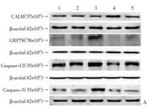
ARG65683 anti-beta Actin antibody WB image
Western blot: Human hepatic stellate cell stained with ARG20531 anti-BiP / GRP78 antibody, ARG54938 anti-Caspase 3 antibody, ARG55123 anti-Calreticulin antibody, ARG55177 anti-Caspase 12 antibody, and ARG65683 anti-beta Actin antibody. Secondary Antibody stained with ARG65351 Goat anti-Rabbit IgG antibody (HRP).
From DAI Linyu et al. Journal of Third Military Medical University (2021), doi: 10-16016-j-1000-5404-202012010, Fig. 4.
-
ARG56491 anti-COX2 antibody WB image
Western blot: Mouse stomach stained with ARG56491 anti-COX2 antibody.
From Zhu M et al. Foods (2025), doi: 10.3390/foods14091600, Fig. 4H.
-
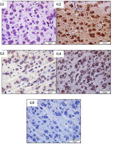
ARG56509 anti-iNOS antibody IHC-P image
Immunohistochemistry: Rat Brain stained with ARG56509 anti-iNOS antibody at 1:100 dilution.
From Abrar Roshdy Abouelkeir et al. European Chemical Bulletin,(2023) doi: 10.31838/ecb/2023.12.1.470, Fig. 6.
-
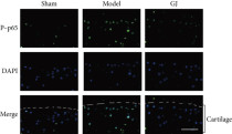
ARG51518 anti-NFkB p65 phospho (Ser536) antibody IHC-P image
Immunohistochemistry: Rat femoral head stained with ARG51518 anti-NFkB p65 phospho (Ser536) antibody at 1:300 dilution.
From Huihui Xu et al. Apoptosis. (2023), doi: 10.1007/s10495-023-01860-2, Fig. 6A.
-
ARG65683 anti-beta Actin antibody WB image
Western blot: Mouse hippocampus stained with ARG22294 anti-SOD1 antibody , ARG54937 anti-SOD2 antibody and ARG65683 anti-beta Actin antibody.
From Zihao Xia et al. International Journal o f Molecular Sciences (2022), doi: 10.3390/ijms23126463, Fig. 6C.
-
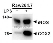
ARG56491 anti-COX2 antibody WB image
Western blot: 20 µg of Raw264.7 cells untreated or treated with LPS. The blots were stained with ARG55060 anti-iNOS antibody at 1:500 dilution and ARG56491 anti-COX2 antibody at 1:200 dilution.
-
ARG56509 anti-iNOS antibody ICC/IF image (Customer's Feedback)
Immunofluorescence: RAW264.7 cells were fixed with 4% paraformaldehyde for 15 min at RT, permeabilized with 0.1% Triton X-100 then blocked with 2% albumin for 60 min at RT. Cells were stained with ARG56509 anti-iNOS antibody (green) at 4°C. DAPI (blue) was used as the nuclear counter stain.
-
ARG51518 anti-NFkB p65 phospho (Ser536) antibody IHC-P image
Immunohistochemistry: Mouse tibial cartilage stained with ARG51518 anti-NFkB p65 phospho (Ser536) antibody.
From Congzi Wu et al. Biomed Res Int. (2022), doi: 10.1155/2022/9230784, Fig. 6. c.
-
ARG65683 anti-beta Actin antibody WB image
Western blot: C2C12 stained with ARG65683 anti-beta Actin antibody.
From Lin YH et al. Biomolecules (2021), doi: 10.3390/biom11111583, Fig. 1. C.
-
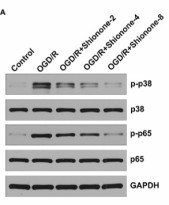
ARG51518 anti-NFkB p65 phospho (Ser536) antibody WB image
Western blot: SH-SY5Y stained with ARG51850 anti-p38 MAPK phospho (Thr180 / Tyr182) antibody, ARG55258 anti-p38 MAPK antibody, ARG51518 anti-NFkB p65 phospho (Ser536) antibody, and ARG57479 anti-NFkB p65 antibody.
From Zhou X et al. J Cardiothorac Surg (2024), doi: 10.1186/s13019-024-02938-x, Fig. 4. A.
-
ARG65683 anti-beta Actin antibody WB image
Western blot: 20 µg of HeLa, Mouse brain and Rat brain lysates stained with ARG65683 anti-beta Actin antibody at 1:10000 dilution.
-
ARG65683 anti-beta Actin antibody WB image
Western blot: 30 µg of 293T lysate stained with ARG65683 anti-beta Actin antibody at 1:3000 dilution.
-
ARG65683 anti-beta Actin antibody WB image
Western blot: 30 µg of 1) Rat brain, and 2) Mouse liver lysate stained with ARG65683 anti-beta Actin antibody at 1:3000 dilution.
-
ARG56509 anti-iNOS antibody WB image
Western blot: Rat Aortic stained with ARG56509 anti-iNOS antibody at 1:1000 dilution.
From Wahid Shah et al. Sci Rep. (2023), doi: 10.1038/s41598-023-43786-4, Fig. 2. C.
-
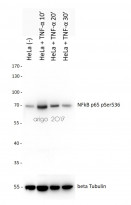
ARG51518 anti-NFkB p65 phospho (Ser536) antibody WB image
Western blot: 20 µg of HeLa cells untreated or treated with TNF-alpha at 10, 20 or 30 min. The blots were stained with ARG51518 anti-NFkB p65 phospho (Ser536) antibody at 1:500 dilution.
-
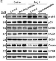
ARG51518 anti-NFkB p65 phospho (Ser536) antibody WB image
Western blot: Mouse heart stained with ARG51518 anti-NFkB p65 phospho (Ser536) antibody.
From Zhang J et al. Frontiers in Pharmacology (2022), doi: 10.3389/fphar.2022.890202, Fig. 4. E.
-
ARG56509 anti-iNOS antibody IHC-P image
Immunohistochemistry: Paraffin-embedded Human pancreatic ductal adenocarcinoma stained with ARG56509 anti-iNOS antibody.
-
ARG56509 anti-iNOS antibody WB image
Western blot: Raw264.7 cells untreated or treated with LPS. 20 µg of cell lysates stained with ARG56509 anti-iNOS antibody at 1:400 dilution.
-
ARG51518 anti-NFkB p65 phospho (Ser536) antibody IHC-P image
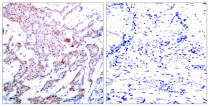
Immunohistochemistry: Paraffin-embedded Human breast carcinoma tissue stained with ARG51518 anti-NFkB p65 phospho (Ser536) antibody (left) or the same antibody preincubated with blocking peptide (right).
-
ARG51518 anti-NFkB p65 phospho (Ser536) antibody ICC/IF image
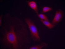
Immunofluorescence: methanol-fixed HeLa cells stained with ARG51518 anti-NFkB p65 phospho (Ser536) antibody.
-
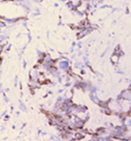
ARG51518 anti-NFkB p65 phospho (Ser536) antibody IHC-P image
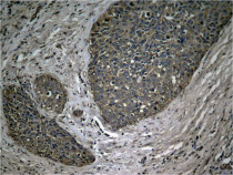
Immunohistochemistry: Paraffin-embedded Human breast carcinoma tissue stained with ARG51518 anti-NFkB p65 phospho (Ser536) antibody.
-
ARG51518 anti-NFkB p65 phospho (Ser536) antibody IHC-P image
Immunohistochemistry: Paraffin-embedded Human Lung carcinoma tissue stained with ARG51518 anti-NFkB p65 phospho (Ser536) antibody.
-
ARG51518 anti-NFkB p65 phospho (Ser536) antibody ICC/IF image
Immunofluorescence: methanol-fixed MEF cells stained with ARG51518 anti-NFkB p65 phospho (Ser536) antibody.
-
ARG65683 anti-beta Actin antibody IHC-P image
Immunohistochemistry: Human ovary tissue stained with ARG65683 anti-beta Actin antibody at 1:200 dilution.
-
ARG56491 anti-COX2 antibody WB image
Western blot: 20 µg of HeLa cell lysate stained with ARG56491 anti-COX2 antibody at 1:200 dilution.
-
ARG65351 Goat anti-Rabbit IgG antibody (HRP) WB image
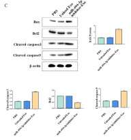
Western blot: Gastric cancer cells stained with ARG66247 anti-Bax antibody, ARG55188 anti-Bcl 2 antibody, ARG57512 anti-Caspase 3 (cleaved) antibody and ARG62346 anti-beta Actin antibody [BA3R].
Secondary Antibody stained with ARG65351 Goat anti-Rabbit IgG antibody (HRP).From Limin Zhang et al. Heliyon (2024), doi: 10.1016/j.heliyon.2024.e30803, Fig. 4. C.
-
ARG65351 Goat anti-Rabbit IgG antibody (HRP) WB image
Western blot: Rat placental stained with ARG57589 anti-MTNR1A antibody at 1:1000 dilution, ARG65351 Goat anti-Rabbit IgG antibody (HRP) at 1:5000 dilution.
From Jinzhi Li et al. J Reprod Immunol. (2023), doi: 10.1016/j.jri.2023.104166, Fig. 2.B.
文献引用
