ARG30131
Postsynaptic Receptor Antibody Panel (NMDAR2A, NMDAR2B, GluR1)
Immune System antibody; Neuroscience antibody
内含物
| 货号 | 内含物名称 | 宿主克隆性 | 反应 | 应用 | 包装 |
|---|---|---|---|---|---|
| ARG52358 | anti-NMDAR2A antibody | Rabbit pAb | Ms, Rat, Rb | IHC-Fr, WB | 50 μl |
| ARG52363 | anti-NMDAR2B antibody | Rabbit pAb | Ms, Rat | WB | 50 μl |
| ARG52314 | anti-GluR1 antibody [RH95] | Mouse mAb | Ms, Rat | WB | 50 μl |
| ARG65350 | Goat anti-Mouse IgG antibody (HRP) | Goat pAb | Ms | ELISA, IHC-P, WB | 50 μl |
| ARG65351 | Goat anti-Rabbit IgG antibody (HRP) | Goat pAb | Rb | ELISA, IHC-P, WB | 50 μl |
概述
| 产品描述 | Synaptic plasticity, the variable efficacy of neurotransmission at synapses, is thought to form the memories. Glutaminergic synapses mediate virtually all excitatory neurotransmission in mammalian brains. Glutamate released from presynaptic terminals activates several types of glutamate-gated ion channels on postsynaptic membranes, including a-amino-3-hydroxy-5-methyl-4-isoxazolepropionic acid (AMPA) receptors, consist of subunits GluR1-GluR4, and N-methyl-D-aspartate (NMDA) receptors. Changes in AMPA receptors at the postsynaptic membrane cause changes in synaptic strength, the best-characterized forms of which are long-term potentiation (LTP) and long-term depression (LTD). This antibody panel could be used to investigate the ratio of NMDAR to AMPAR or the change of AMPAR on the postsynaptic membrane. During the LTP, the AMPARs increase expression on the membrane of post-synapse, and the ratio of NMDAR to AMPAR would be higher than the pre-LTP. In construct, LTD decreases the AMPARs expression, and the ratio would be lower than pre-LTD. |
|---|---|
| 靶点名称 | Postsynaptic Receptor |
| 別名 | Postsynaptic Receptor antibody; GluR1 antibody; NMDAR2A antibody; NMDAR2B antibody |
属性
| 存放说明 | For continuous use, store undiluted antibody at 2-8°C for up to a week. For long-term storage, aliquot and store at -20°C or below. Storage in frost free freezers is not recommended. Avoid repeated freeze/thaw cycles. Suggest spin the vial prior to opening. The antibody solution should be gently mixed before use. |
|---|---|
| 注意事项 | For laboratory research only, not for drug, diagnostic or other use. |
生物信息
| 全名 | Antibody Panel for Postsynaptic Receptor (NMDAR2A, NMDAR2B, GluR1) |
|---|---|
| 产品亮点 | Related Product: anti-NMDAR2A antibody; anti-NMDAR2B antibody; anti-GluR1 antibody; |
| 研究领域 | Immune System antibody; Neuroscience antibody |
检测图片 (10) Click the Picture to Zoom In
-

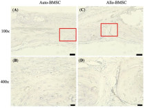
ARG65351 Goat anti-Rabbit IgG antibody (HRP) IHC-P image
From Yu-Qian Song et al. J Mol Med (Berl) (2022), doi: 10.1007/s00109-021-02165-0, Fig. 5.c.
-

ARG65351 Goat anti-Rabbit IgG antibody (HRP) WB image
Western blot: HK2 stained with ARG66421 anti-CXCL12 / SDF1 antibody [SQab18123] and ARG10112 anti-GAPDH antibody [6C5] and secondary antibody used ARG65351 Goat anti-Rabbit IgG antibody (HRP).
From Zhou Y et al. Central European Journal of Immunology (2022), doi: 10.5114/ceji.2022.115628, Fig. 2. E.
-
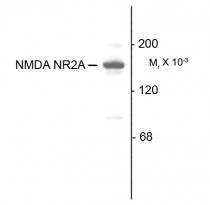
ARG52358 anti-NMDAR2A antibody WB image
Western Blot: 10 µg of Rat hippocampal (Hipp) lysate stained with ARG52358 anti-NMDAR2A antibody showing specific immunolabeling of the ~180 kDa NR2A subunit of the NMDA receptor.
-
ARG52314 anti-GluR1 antibody [RH95] WB image
Western Blot: rat hippocampal lysate showing specific immunolabeling of the ~105k GluR1 protein stained with GluR1 antibody (ARG52314).
-
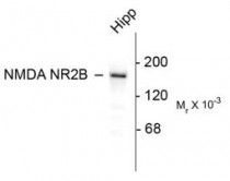
ARG52363 anti-NMDAR2B antibody WB image
Western blot: 10 ug of rat hippocampal (Hipp) lysate stained with ARG52363 anti-NMDAR2B antibody showing specific immunolabeling of the ~180k NR2B subunit of the NMDA receptor.
-
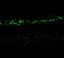
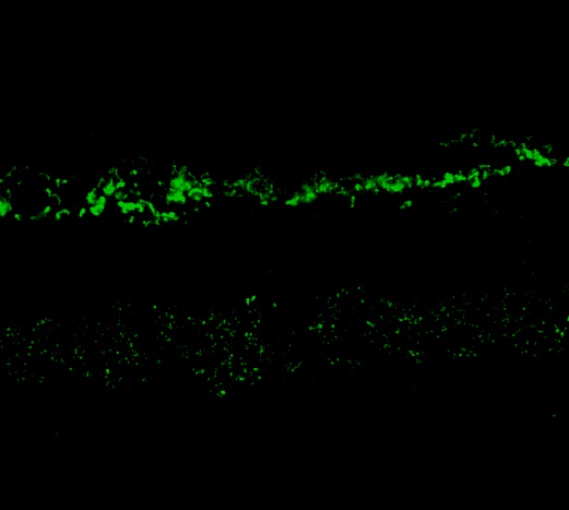
ARG52358 anti-NMDAR2A antibody IHC-Fr image
Immunohistochemistry: Rabbit retina stained with ARG52358 anti-NMDAR2A antibody showing NR2A in the rod and cone photoreceptors in the outer plexiform layer as well as the entire inner plexiform layer.
-
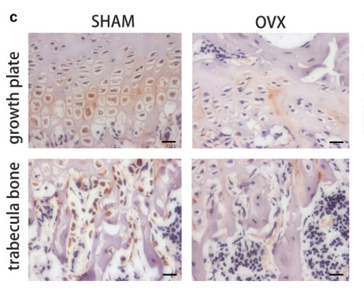
ARG65350 Goat anti-Mouse IgG antibody (HRP) IHC-P image
From Cheng-Feng Chu et al. J Pers Med. (2021), doi: 10.3390/jpm11121326, Fig. 6.
-
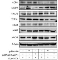
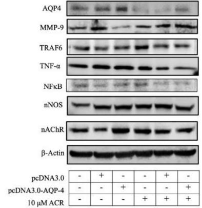
ARG65350 Goat anti-Mouse IgG antibody (HRP) WB image
Western blot: Neuro-2a stained with ARG22361 anti-Aquaporin 4 antibody , ARG40638 anti-MMP9 antibody and ARG65350 Goat anti-Mouse IgG antibody (HRP).
From Hung CY et al. Molecules (2022), doi: 10.3390/molecules27031066, Fig. 6A.
-
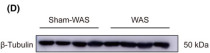
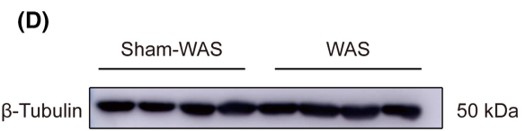
ARG65350 Goat anti-Mouse IgG antibody (HRP) WB image
Western blot: Rat basolateral amygdala stained with ARG62347 anti-beta Tubulin antibody [BT7R] at 1:1000 dilution, ARG65350 Goat anti-Mouse IgG antibody (HRP) at 1:5000 dilution.
From Guang-Bing Duan et al. CNS Neurosci Ther. (2024), doi: 10.1111/cns.14611, Fig. 4.D.
-

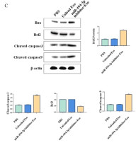
ARG65351 Goat anti-Rabbit IgG antibody (HRP) WB image
Western blot: Gastric cancer cells stained with ARG66247 anti-Bax antibody, ARG55188 anti-Bcl 2 antibody, ARG57512 anti-Caspase 3 (cleaved) antibody and ARG62346 anti-beta Actin antibody [BA3R].
Secondary Antibody stained with ARG65351 Goat anti-Rabbit IgG antibody (HRP).From Limin Zhang et al. Heliyon (2024), doi: 10.1016/j.heliyon.2024.e30803, Fig. 4. C.


文献引用
A New Way to Engineer Cell Sheets for Articular Cartilage Regeneration
ARG65350; ICC/IF / Rabbit


Soybean Trypsin Inhibitor Possesses Potency Against SARS-CoV-2 Infection by Blocking the Host Cell Surface Receptors ACE2, TMPRSS2, and CD147
ARG65351; WB /
Progressively diminished estrogen signaling concordant with increased fibrosis in ectopic endometrium
ARG65351; WB /
Expression and correlation of Surfeit 4 gene in esophageal squamous cell carcinoma
ARG65350; WB /
Environmental acidification drives inter-organ energy mobilization to enhance reproductive performance in medaka (Oryzias latipes)
ARG65350; WB /
KDF1 Promoted Proliferation, Migration and Invasion of Lung Adenocarcinoma Cells through Activating STAT3 and AKT Pathway
ARG65350: WB /


