ARG30275
Pro-apoptotic Bcl2 protein Antibody Panel (BAX, BAK, Bid)
内含物
| 货号 | 内含物名称 | 宿主克隆性 | 反应 | 应用 | 包装 |
|---|---|---|---|---|---|
| ARG54941 | anti-Bax antibody | Rabbit pAb | Hu, Ms | ICC/IF, IP, WB | 50 μg |
| ARG62367 | anti-Bak antibody | Rabbit pAb | Hu, Ms, Hm | IHC-P, WB | 100 μl |
| ARG65625 | anti-Bid antibody | Rabbit pAb | Hu | IHC-P, WB | 50 μl |
| ARG65351 | Goat anti-Rabbit IgG antibody (HRP) | Goat pAb | Rb | ELISA, IHC-P, WB | 50 μl |
概述
| 产品描述 | A variety of physiological death signals trigger the genetically programmed pathway of apoptosis which is manifested in two major execution programs downstream of the death signal: the caspase pathway and organelle dysfunction. The BCL-2 family of proteins is known as an important gatekeeper to the apoptotic response. This group of structurally related proteins comprises pro-apoptotic and anti-apoptotic members that interact with one another. The pro-apoptotic factors, including BAX, BAK or BID promotes pore-forming effectors such that Cytochrome-C is released from the mitochondria and kick-start the caspase cleavage cascades. They, together with their opposing partner, BCL2, have become targets for cancer therapeutic interventions. Gross et al. 1999. Genes & Dev 13:1899-1911 |
|---|---|
| 靶点名称 | Pro-apoptotic Bcl2 protein |
| 別名 | Pro-apoptotic Bcl2 protein antibody; Bax antibody; Bak antibody; Bid antibody |
属性
| 存放说明 | For continuous use, store undiluted antibody at 2-8°C for up to a week. For long-term storage, aliquot and store at -20°C or below. Storage in frost free freezers is not recommended. Avoid repeated freeze/thaw cycles. Suggest spin the vial prior to opening. The antibody solution should be gently mixed before use. |
|---|---|
| 注意事项 | For laboratory research only, not for drug, diagnostic or other use. |
生物信息
| 全名 | Antibody Panel for Pro-apoptotic Bcl2 protein (BAX, BAK, Bid) |
|---|---|
| 产品亮点 | Related Product: anti-Bax antibody; anti-Bak antibody; anti-Bid antibody; |
检测图片 (11) Click the Picture to Zoom In
-

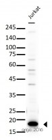
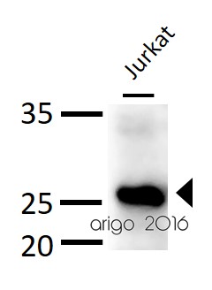
ARG62367 anti-Bak antibody WB image
Western blot: 30 µg of Jurkat cell lysate stained with ARG62367 anti-Bak antibody at 1:500 dilution.
-

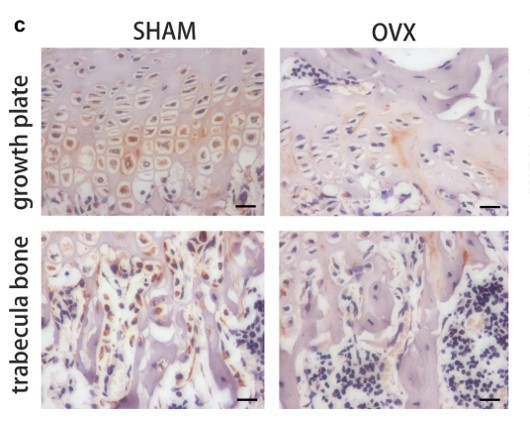
ARG65351 Goat anti-Rabbit IgG antibody (HRP) IHC-P image
From Yu-Qian Song et al. J Mol Med (Berl) (2022), doi: 10.1007/s00109-021-02165-0, Fig. 5.c.
-
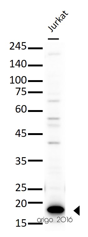
ARG65625 anti-Bid antibody WB image
Western blot: 30 µg of Jurkat cell lysate stained with ARG65625 anti-Bid antibody at 1:500 dilution.
-

ARG65351 Goat anti-Rabbit IgG antibody (HRP) WB image
Western blot: HK2 stained with ARG66421 anti-CXCL12 / SDF1 antibody [SQab18123] and ARG10112 anti-GAPDH antibody [6C5] and secondary antibody used ARG65351 Goat anti-Rabbit IgG antibody (HRP).
From Zhou Y et al. Central European Journal of Immunology (2022), doi: 10.5114/ceji.2022.115628, Fig. 2. E.
-

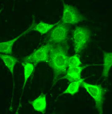
ARG54941 anti-Bax antibody ICC/IF image
Immunofluorescence: NIH/3T3 cells stained with ARG54941 anti-Bax antibody at 1:100 dilution.
-

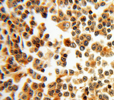
ARG54941 anti-Bax antibody IHC-P image
Immunohistochemistry: paraffin-embedded Lymphoma stained with ARG54941 anti-Bax antibody.
-
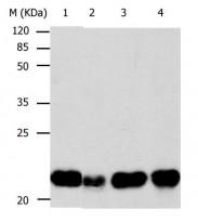
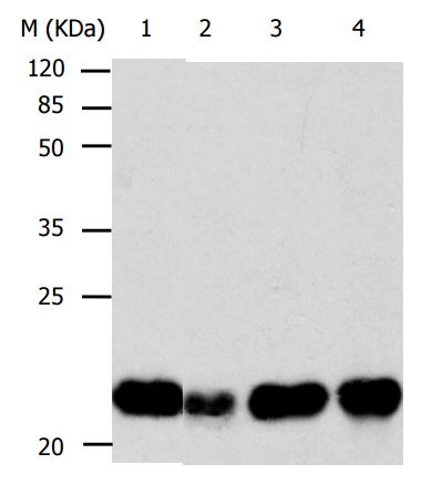
ARG54941 anti-Bax antibody WB image
Western blot: 30 µg of 1) HeLa, 2) HepG2, 3) 293T, and 4) Mouse ovary lysate stained with ARG54941 anti-Bax antibody at 1:500 dilution.
-
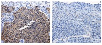
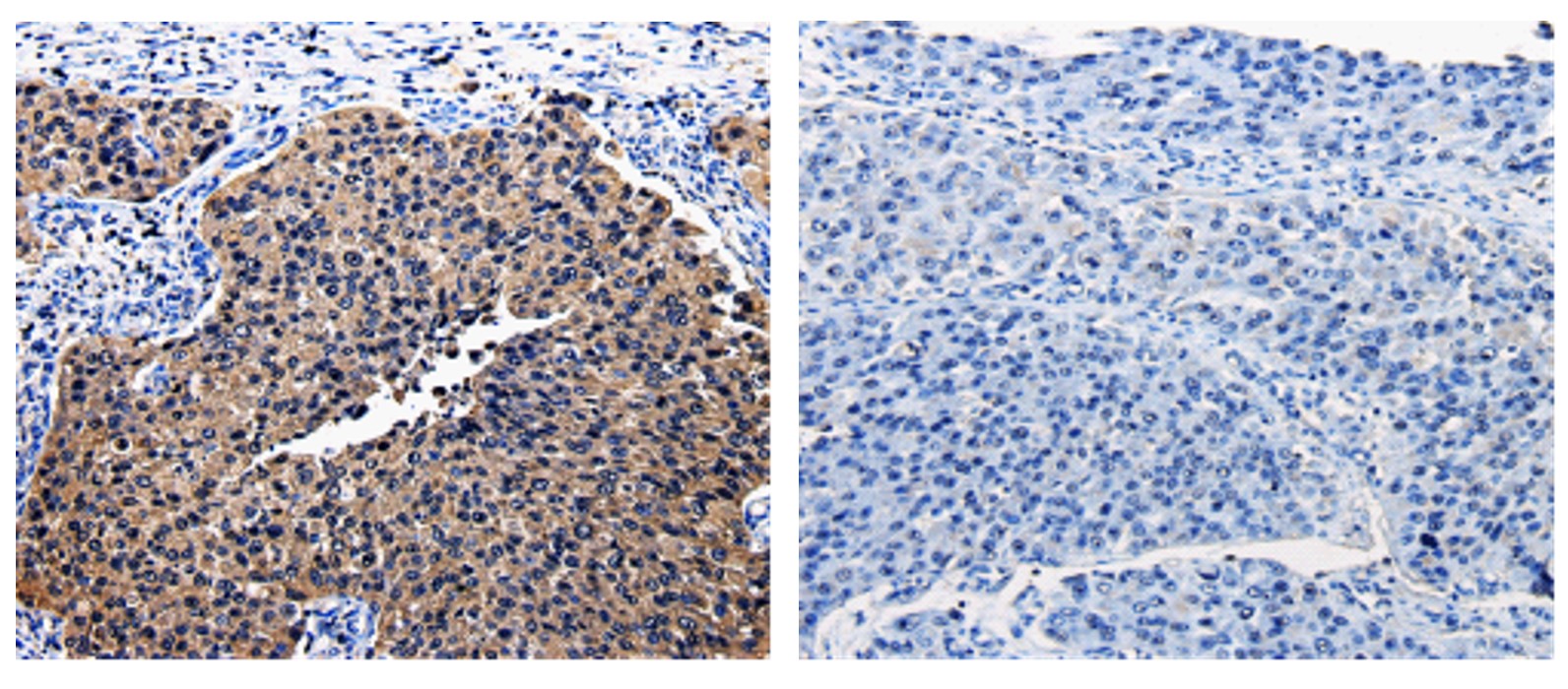
ARG65625 anti-Bid antibody IHC-P image
Immunohistochemistry: Paraffin-embedded Human renal cancer tissue stained with ARG65625 anti-Bid antibody (left) at 1:25 dilution, or the same antibody preincubated with antigen (right). (Original magnification: X200)
-
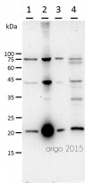
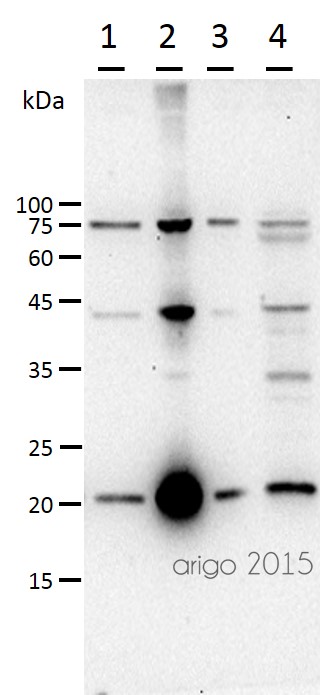
ARG65625 anti-Bid antibody WB image
Western Blot: 40 μg of lysates from: 1) Human fetal brain tissue, 2) Human liver tissue, 3) HT29 cells, 4) HepG2 cells stained with ARG65625 anti-Bid antibody at 1/260 dilution. Exposure time: 10 seconds.
-

ARG65351 Goat anti-Rabbit IgG antibody (HRP) WB image
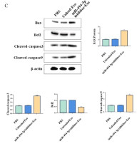
Western blot: Gastric cancer cells stained with ARG66247 anti-Bax antibody, ARG55188 anti-Bcl 2 antibody, ARG57512 anti-Caspase 3 (cleaved) antibody and ARG62346 anti-beta Actin antibody [BA3R].
Secondary Antibody stained with ARG65351 Goat anti-Rabbit IgG antibody (HRP).From Limin Zhang et al. Heliyon (2024), doi: 10.1016/j.heliyon.2024.e30803, Fig. 4. C.
-

ARG65351 Goat anti-Rabbit IgG antibody (HRP) WB image
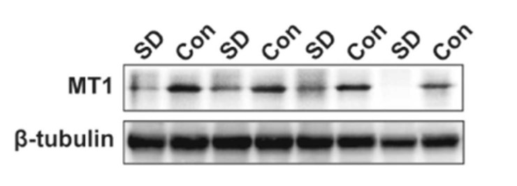
Western blot: Rat placental stained with ARG57589 anti-MTNR1A antibody at 1:1000 dilution, ARG65351 Goat anti-Rabbit IgG antibody (HRP) at 1:5000 dilution.
From Jinzhi Li et al. J Reprod Immunol. (2023), doi: 10.1016/j.jri.2023.104166, Fig. 2.B.

文献引用
Soybean Trypsin Inhibitor Possesses Potency Against SARS-CoV-2 Infection by Blocking the Host Cell Surface Receptors ACE2, TMPRSS2, and CD147
ARG65351; WB /


Progressively diminished estrogen signaling concordant with increased fibrosis in ectopic endometrium
ARG65351; WB /


