ARG30227
Phospho ATF2 Antibody Panel (Total, pT69, pT71)
Gene Regulation antibody; Immune System antibody
内含物
| 货号 | 内含物名称 | 宿主克隆性 | 反应 | 应用 | 包装 |
|---|---|---|---|---|---|
| ARG51535 | anti-ATF2 phospho (Thr71 or 53) antibody | Rabbit pAb | Hu, Ms, Rat | IHC-P, WB | 50 μl |
| ARG51534 | anti-ATF2 phospho (Thr69 or 51) antibody | Rabbit pAb | Hu, Ms, Rat | ICC/IF, IHC-P, WB | 50 μl |
| ARG63122 | anti-ATF2 antibody | Goat pAb | Hu | WB | 50 μg |
| ARG65351 | Goat anti-Rabbit IgG antibody (HRP) | Goat pAb | Rb | ELISA, IHC-P, WB | 50 μl |
| ARG65352 | Donkey anti-Goat IgG antibody (HRP) | Donkey pAb | Goat | ELISA, IHC-P, WB | 50 μl |
概述
| 产品描述 | The activating transcription factor (ATF) and cAMP-response-element binding (CREB) families of transcription factors comprise 16 members of the activator protein 1 (AP1) transcription factor superfamily. ATF and CREB proteins can homodimerize or heterodimerize with members of the Jun, Fos or Maf transcription factor families to form complexes that regulate diverse cellular functions, such as stress responses, embryonic development, disease development and cell death. ATF2 requires phosphorylation by Jun N-terminal kinase (JNK), p38 (MAPK14), or extracellular-signal-regulated kinase 1 (ERK1) at Thr69 and Thr71 residues in order to be transcriptionally active. Lau and Ronai. 2012. J Cell Sci 125:2815 Morton et al. 2004. FEBS Lett 572:177-83 Waas et al. 2001. J Biol Chem 276(8):5676 |
|---|---|
| 靶点名称 | ATF2 |
| 別名 | Phospho ATF2 antibody; Phospho Activating transcription factor 2 antibody; ATF2 phospho (Thr69 or 51) antibody; ATF2 phospho (Thr71 or 53) antibody; ATF2 antibody |
属性
| 存放说明 | For continuous use, store undiluted antibody at 2-8°C for up to a week. For long-term storage, aliquot and store at -20°C or below. Storage in frost free freezers is not recommended. Avoid repeated freeze/thaw cycles. Suggest spin the vial prior to opening. The antibody solution should be gently mixed before use. |
|---|---|
| 注意事项 | For laboratory research only, not for drug, diagnostic or other use. |
生物信息
| 全名 | Phospho Activating transcription factor 2 (ATF2) Antibody Panel (Total, pT69, pT71) |
|---|---|
| 产品亮点 | Related Product: anti-ATF2 antibody; |
| 研究领域 | Gene Regulation antibody; Immune System antibody |
检测图片 (12) Click the Picture to Zoom In
-

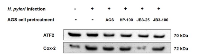
ARG63122 anti-ATF2 antibody WB image
Western blot: AGS cells stained with ARG56421 anti-COX2 antibody, ARG63122 anti-ATF2 antibody and ARG65352 Donkey anti-Goat IgG antibody (HRP).
From Anh Duy Do et al. Front Immunol. (2022), doi: 10.3389/fimmu.2021.796177, Fig. 5.D.
-

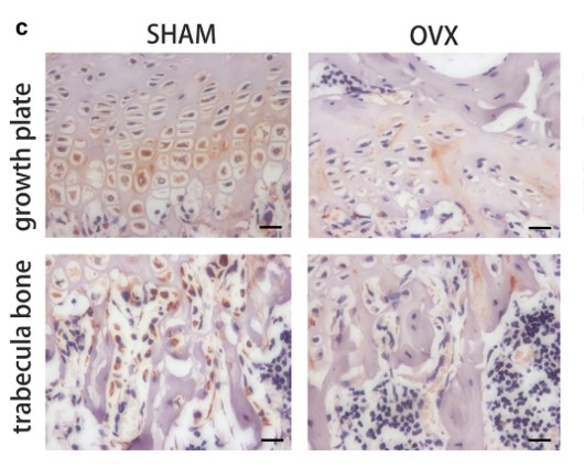
ARG65351 Goat anti-Rabbit IgG antibody (HRP) IHC-P image
From Yu-Qian Song et al. J Mol Med (Berl) (2022), doi: 10.1007/s00109-021-02165-0, Fig. 5.c.
-

ARG65351 Goat anti-Rabbit IgG antibody (HRP) WB image
Western blot: HK2 stained with ARG66421 anti-CXCL12 / SDF1 antibody [SQab18123] and ARG10112 anti-GAPDH antibody [6C5] and secondary antibody used ARG65351 Goat anti-Rabbit IgG antibody (HRP).
From Zhou Y et al. Central European Journal of Immunology (2022), doi: 10.5114/ceji.2022.115628, Fig. 2. E.
-
_210_205.jpg)
ARG65352 Donkey anti-Goat IgG antibody (HRP) WB image
Western blot: Endometrial Stromal cells stained with ARG63922 anti-EP4 prostaglandin Receptor antibody at 1:1000 dilution and ARG65352 Donkey anti-Goat IgG antibody (HRP).
From Qingqing Huang et al. Reprod Med Biol. (2021), doi: 10.1002/rmb2.12423, Fig. 3.C.
-
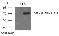
ARG51534 anti-ATF2 phospho (Thr69 or 51) antibody WB image
Western Blot: extracts from 3T3 cells untreated or treated with Anisomycin stained with anti-ATF2 (phospho Thr69 or 51) antibody ARG51534.
-

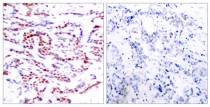
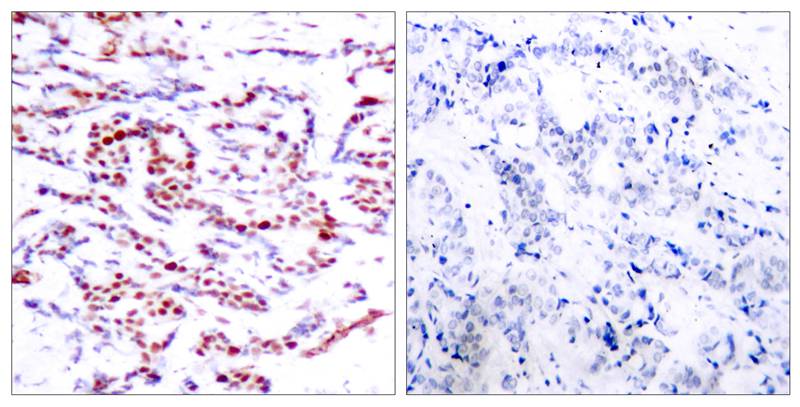
ARG51534 anti-ATF2 phospho (Thr69 or 51) antibody IHC-P image
Immunohistochemistry: paraffin-embedded human breast carcinoma tissue stained with anti-ATF2 (phospho Thr69 or 51) antibody ARG51534 (left) or the same antibody preincubated with blocking peptide (right).
-

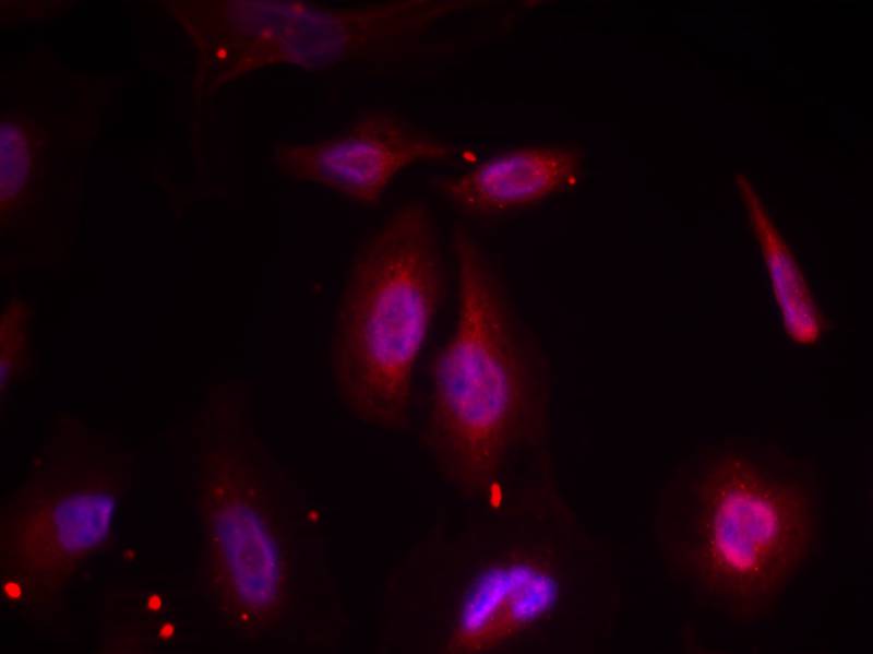
ARG51534 anti-ATF2 phospho (Thr69 or 51) antibody ICC/IF image
Immunofluorescence: methanol-fixed HeLa cells stained with anti-ATF2 (phospho Thr69 or 51) antibody ARG51534.
-
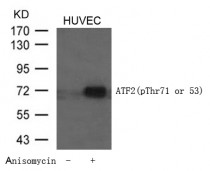
ARG51535 anti-ATF2 phospho (Thr71 or 53) antibody WB image
Western Blot: extracts from HUVEC cells untreated or treated with Anisomycin stained with anti-ATF2 (phospho Thr71 or 53) antibody ARG51535.
-
ARG51535 anti-ATF2 phospho (Thr71 or 53) antibody IHC-P image
Immunohistochemistry: paraffin-embedded human breast carcinoma tissue stained with anti-ATF2 (phospho Thr71 or 53) antibody ARG51535 (left) or the same antibody preincubated with blocking peptide (right).
-
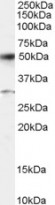
ARG63122 anti-ATF2 antibody WB image
Western Blot: Human Placenta lysate (RIPA buffer, 35 µg total protein per lane) stained with ARG63122 anti-ATF2 antibody at 0.5 µg/ml dilution.
-

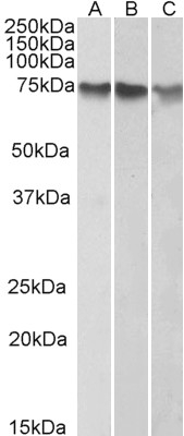
ARG63122 anti-ATF2 antibody WB image
Western Blot: HepG2 (A), K562 (B) and HeLa (C) nuclear lysates (35 µg protein in RIPA buffer) stained with ARG63122 anti-ATF2 antibody at 1 µg/ml dilution.
-

ARG65351 Goat anti-Rabbit IgG antibody (HRP) WB image
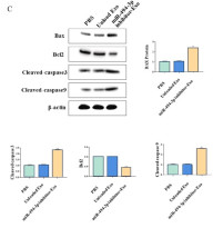
Western blot: Gastric cancer cells stained with ARG66247 anti-Bax antibody, ARG55188 anti-Bcl 2 antibody, ARG57512 anti-Caspase 3 (cleaved) antibody and ARG62346 anti-beta Actin antibody [BA3R].
Secondary Antibody stained with ARG65351 Goat anti-Rabbit IgG antibody (HRP).From Limin Zhang et al. Heliyon (2024), doi: 10.1016/j.heliyon.2024.e30803, Fig. 4. C.

.jpg)


文献引用
Progressively diminished estrogen signaling concordant with increased fibrosis in ectopic endometrium
ARG65351; WB /


Soybean Trypsin Inhibitor Possesses Potency Against SARS-CoV-2 Infection by Blocking the Host Cell Surface Receptors ACE2, TMPRSS2, and CD147
ARG65351; WB /


