ARG30316
ER Marker Antibody Duo
内含物
| 货号 | 内含物名称 | 宿主克隆性 | 反应 | 应用 | 包装 |
|---|---|---|---|---|---|
| ARG55123 | anti-Calreticulin antibody | Rabbit pAb | Hu, Ms, Rat | ICC/IF, IHC-P, WB | 50 μl |
| ARG20531 | anti-BiP / GRP78 antibody | Rabbit pAb | Hu, Ms, Rat, Bov, Dog, Fungi, Hm, Mk, Rb, Xenopus laevis | ICC/IF, IHC-FoFr , IHC-P, IP, WB | 50 μl |
概述
| 产品描述 | Endoplasmic reticulum (ER) is a network of membrane-enclosed tubules and sacs (cisternae) that extends from the nuclear membrane throughout the cytoplasm. The ER lumen is the area enclosed by the ER membrane. ER is responsible for protein synthesis and folding, protein transport into the Golgi apparatus, and lipid metabolism. arigo's ER Marker Antibody Duo comprises an ER membrane marker Calreticulin antibody and an ER lumen marker BiP antibody. It is an excellent solution for labeling ER structures. |
|---|---|
| 靶点名称 | ER Marker |
| 別名 | ER Marker antibody; BiP / GRP78 antibody; Calreticulin antibody |
属性
| 存放说明 | For continuous use, store undiluted antibody at 2-8°C for up to a week. For long-term storage, aliquot and store at -20°C or below. Storage in frost free freezers is not recommended. Avoid repeated freeze/thaw cycles. Suggest spin the vial prior to opening. The antibody solution should be gently mixed before use. |
|---|---|
| 注意事项 | For laboratory research only, not for drug, diagnostic or other use. |
生物信息
| 全名 | Antibody Duo for ER Marker |
|---|---|
| 产品亮点 | Related Product: anti-Calreticulin antibody; anti-BiP / GRP78 antibody; |
检测图片 (11) Click the Picture to Zoom In
-

ARG55123 anti-Calreticulin antibody WB image
Western blot: Human hepatic stellate cell stained with ARG20531 anti-BiP / GRP78 antibody, ARG54938 anti-Caspase 3 antibody, ARG55123 anti-Calreticulin antibody, ARG55177 anti-Caspase 12 antibody, and ARG65683 anti-beta Actin antibody. Secondary Antibody stained with ARG65351 Goat anti-Rabbit IgG antibody (HRP).
From DAI Linyu et al. Journal of Third Military Medical University (2021), doi: 10-16016-j-1000-5404-202012010, Fig. 4.
-
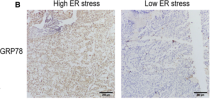
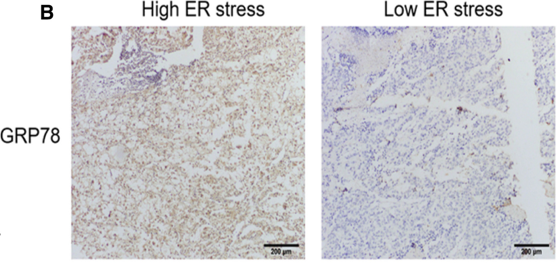
ARG20531 anti-BiP / GRP78 antibody IHC-P image
Immunohistochemistry: Human hepatocellular carcinoma stained with ARG20531 anti-BiP / GRP78 antibody at 1:500 dilution.
From Yue Zhu et al. Cancer Med. (2020), doi: 10.1002/cam4.2977, Fig. 5B.
-
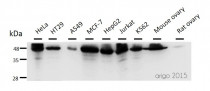
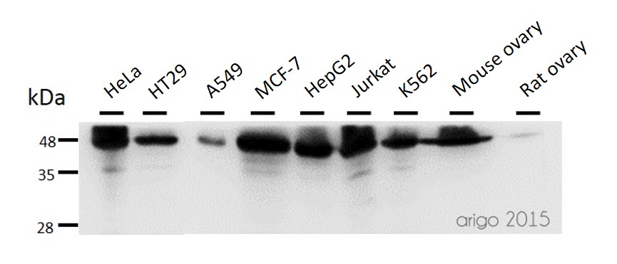
ARG55123 anti-Calreticulin antibody WB image
Western blot: 30 µg of HeLa, HT29, A549, MCF-7, HepG2, Jurkat, K562, Mouse ovary and Rat ovary lysates stained with ARG55123 anti-Calreticulin antibody at 1:500 dilution.
-
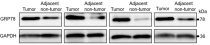
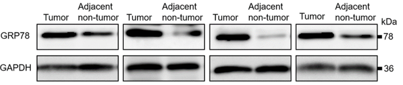
ARG20531 anti-BiP / GRP78 antibody WB image
Western blot: Human hepatocellular carcinoma stained with ARG20531 anti-BiP / GRP78 antibody at 1:500 dilution.
From Yue Zhu et al. Cancer Med. (2020), doi: 10.1002/cam4.2977, Fig. 5D.
-

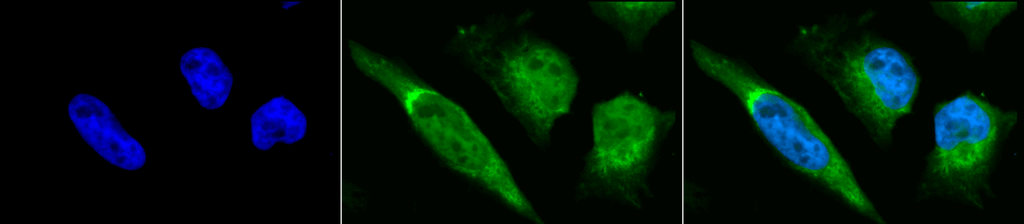
ARG20531 anti-BiP / GRP78 antibody ICC/IF image
Immunofluorescence: Heat Shocked (42°C for 30 min) HeLa cells. Fixation: 2% Formaldehyde for 20 min at RT. Primary Antibody: ARG20531 anti-BiP / GRP78 antibody at 1:100 for 12 hours at 4°C. Secondary Antibody: FITC Goat anti-Rabbit (green) at 1:200 for 2 hours at RT. Counterstain: DAPI (blue) nuclear stain at 1:40000 for 2 hours at RT. Magnification: 100x. Left: DAPI (blue) nuclear stain. Middle: ARG20531 anti-BiP / GRP78 antibody. Right: Composite.
-

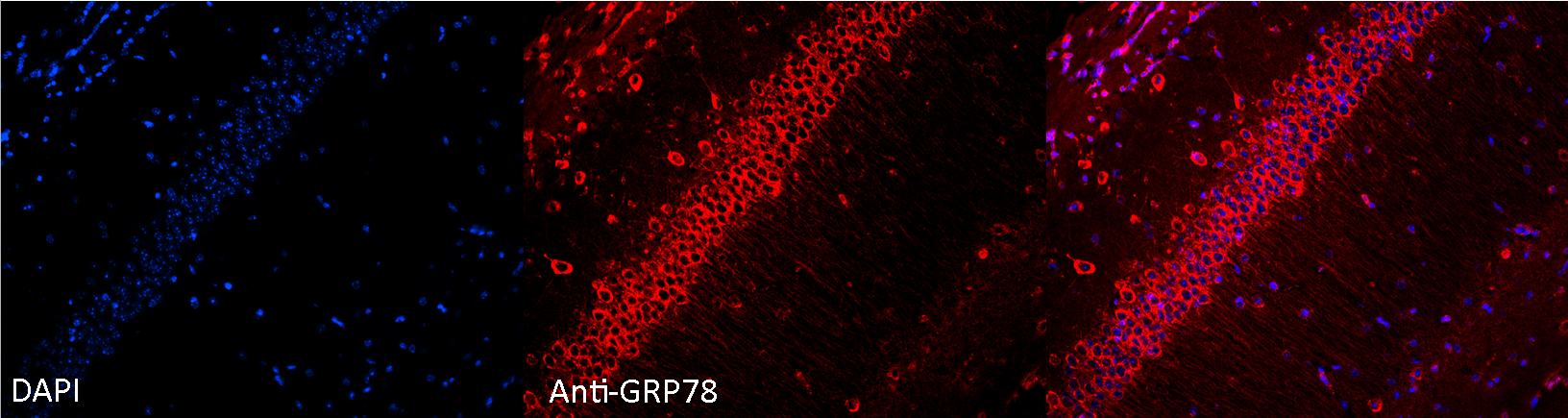
ARG20531 anti-BiP / GRP78 antibody IHC image
Immunohistochemistry: Formaldehdye-fixed and parrafin embedded sections of Mouse hippocampal tissue stained with ARG20531 anti-BiP / GRP78 antibody at 1:100 dilution. Tissue was counterstained using DAPI at a 1:1000 dilution in order to visualize the nuclei in the pyramidal cell layer. Images courtsey of Rachel Reith, NIH/NIMH.
-
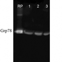
ARG20531 anti-BiP / GRP78 antibody WB image
Western blot: Human, Dog, Mouse cell line lysates stained with ARG20531 anti-BiP / GRP78 antibody at 1:1000 dilution.
-

ARG55123 anti-Calreticulin antibody ICC/IF image
Immunofluorescence: 100% Methanol fixed (RT, 10 min) HeLa cells stained with ARG55123 anti-Calreticulin antibody (red) at 1:20 dilution.
Secondary antibody: ARG21917 Goat anti-Rabbit IgG antibody (TRITC)
-

ARG55123 anti-Calreticulin antibody ICC/IF image
Immunofluorescence: 100% Methanol fixed (RT, 10 min) HeLa cells stained with ARG55123 anti-Calreticulin antibody at 1:20 dilution. Left: primary antibody (red). Middle: DAPI (blue). Right: Merge.
Secondary antibody: ARG21917 Goat anti-Rabbit IgG antibody (TRITC)
-

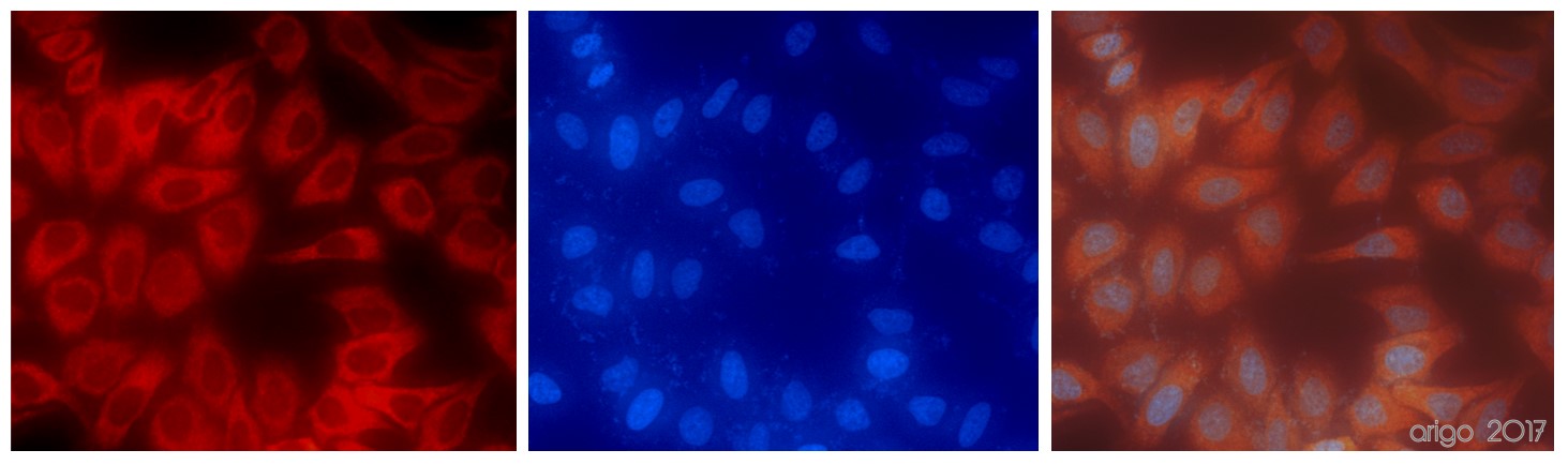
ARG20531 anti-BiP / GRP78 antibody ICC/IF image
Immunofluorescence: Heat Shocked (42°C for 30 min) HeLa cells. Fixation: 2% Formaldehyde for 20 min at RT. Primary Antibody: ARG20531 anti-BiP / GRP78 antibody at 1:100 for 12 hours at 4°C. Secondary Antibody: APC Goat anti-Rabbit (red) at 1:200 for 2 hours at RT. Counterstain: DAPI (blue) nuclear stain at 1:40000 for 2 hours at RT. Magnification: 20x. Left: DAPI (blue) nuclear stain. Middle: ARG20531 anti-BiP / GRP78 antibody. Right: Composite.
-

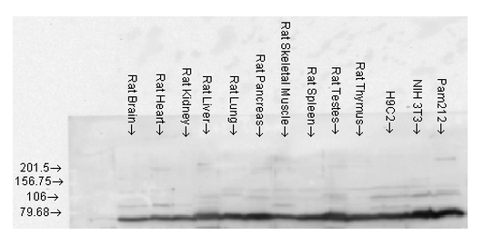
ARG20531 anti-BiP / GRP78 antibody WB image
Western blot: Rat brain, heart, kidney, liver, lung, pancreas, skeletal muscle, spleen, testes, thymus, H9C2 cell, Mouse NIH 3T3 and Pam212 cell lysates stained with ARG20531 anti-BiP / GRP78 antibody.


文献引用
Melatonin Reduces Apoptosis Resistance of Hepatocellular Carcinoma Cells by Inhibiting BMAL1
ARG20531:IHC-P / Human


Down-regulation of calreticulin promotes apoptosis in hepatic stellate cells.
ARG20531, ARG55123: WB / Human
Sp2 promotes invasion and metastasis of hepatocellular carcinoma by targeting TRIB3 protein.
ARG20531: WB, IHC-P / Human
Resveratrol protects against CIH-induced myocardial injury by targeting Nrf2 and blocking NLRP3 inflammasome activation.
ARG20531: WB / Rat
Hydrogen and Oxygen Mixture to Improve Cardiac Dysfunction and Myocardial Pathological Changes Induced by Intermittent Hypoxia in Rats.
ARG20531: WB / Rat
Regulation of P300 and HDAC1 on endoplasmic reticulum stress in isoniazid-induced HL-7702 hepatocyte injury.
ARG20531: WB / Human
Interplay between Redox Signaling, Oxidative Stress, and Unfolded Protein Response (UPR) in Pathogenesis of Human Diseases.
ARG20531: WB / Rat


