ARG30322
Myofibroblast Differentiation Antibody Duo
Cancer antibody; Cell Biology and Cellular Response antibody; Controls and Markers antibody; Developmental Biology antibody; Neuroscience antibody; Signaling Transduction antibody
内含物
| 货号 | 内含物名称 | 宿主克隆性 | 反应 | 应用 | 包装 |
|---|---|---|---|---|---|
| ARG66199 | anti-Vimentin antibody [SQab1721] | Rabbit mAb | Hu, Ms | FACS, ICC/IF, IHC-P, IHC-Fr, IP, WB | 50 μl |
| ARG66381 | anti-alpha smooth muscle Actin antibody [SQab18108] | Rabbit mAb | AGMK, Bov, Ctl, Dog, Hu, Ms, Pig, Rat, Zfsh | FACS, ICC/IF, IHC-P, IHC-Fr, WB | 50 μl |
概述
| 产品描述 | Myofibroblast differentiation is a highly regulated process and plays a critical role in tissue repair, wound healing and chronic fibrosis. This antibody duo comprises fibroblast marker Vimentin antibody and myofibroblast marker smooth muscle actin antibody. It is ideal for the research of fibroblast-to-myofibroblast differentiation. Related news: New antibody panels for Myofibroblasts and CAFs New antibody panels and duos for Tumor immune microenvironment Anti-SerpinB9 therapy, a new strategy for cancer therapy |
|---|---|
| 靶点名称 | Myofibroblast Differentiation |
| 別名 | CAF Marker antibody; Cancer-associated fibroblast Marker antibody; Vimentin antibody; alpha smooth muscle Actin antibody |
属性
| 存放说明 | For continuous use, store undiluted antibody at 2-8°C for up to a week. For long-term storage, aliquot and store at -20°C or below. Storage in frost free freezers is not recommended. Avoid repeated freeze/thaw cycles. Suggest spin the vial prior to opening. The antibody solution should be gently mixed before use. |
|---|---|
| 注意事项 | For laboratory research only, not for drug, diagnostic or other use. |
生物信息
| 全名 | Cancer-associated fibroblast (CAF) Marker Antibody Duo |
|---|---|
| 产品亮点 | Related Product: anti-Vimentin antibody; anti-alpha smooth muscle Actin antibody; |
| 研究领域 | Cancer antibody; Cell Biology and Cellular Response antibody; Controls and Markers antibody; Developmental Biology antibody; Neuroscience antibody; Signaling Transduction antibody |
检测图片 (23) Click the Picture to Zoom In
-
ARG66199 anti-Vimentin antibody [SQab1721] IHC-P image
Immunohistochemistry: C33A stained with ARG66162 anti-Fibronectin antibody , ARG22587 anti-N Cadherin antibody [13A9], ARG66199 anti-Vimentin antibody [SQab1721], ARG59198 anti-Laminin antibody and ARG66195 anti-E Cadherin antibody [SQab1717] .
From Ya-Hui Chen et al. Cancers (Basel)- (2022), doi: 10.3390/cancers14071824, Fig. 6. E.
-
ARG66199 anti-Vimentin antibody [SQab1721] ICC/IF image
Immunofluorescence: Human keloid stained with ARG66199 anti-Vimentin antibody [SQab1721].
From Xiaoqian Li et al. preprint. (2024), doi: 10.21203/rs.3.rs-4780437/v1, Fig. 1. C.
-
ARG66381 anti-alpha smooth muscle Actin antibody [SQab18108] ICC-IF image
Immunofluorescence: Mouse kidney stained with ARG66381 anti-alpha smooth muscle Actin antibody [SQab18108].
From Pengfei Zhu et al. Pharmaceuticals (Basel)- (2022), doi: 10.3390/ph15040434, Fig. 1. A.
-
ARG66199 anti-Vimentin antibody [SQab1721] IHC-P image
Immunohistochemistry: Mouse myometrium stained with ARG66199 anti-Vimentin antibody [SQab1721].
From Xi Wang et al. Biomedicines (2022), doi: 10.3390/biomedicines10061218, Fig. 4. B.
-
ARG66199 anti-Vimentin antibody [SQab1721] IHC-Fr image
Immunohistochemistry: Frozen mouse heart stained with ARG66199 anti-Vimentin antibody [SQab1721].
From Yi-Chao Hsu et al. Cells (2022), doi: 10.3390/cells11010121, Fig. 5. A, 5. E, 5. I,5. M, and 5. Q.
-
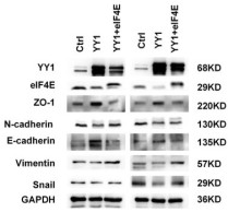
ARG66199 anti-Vimentin antibody [SQab1721] WB image
Western blot: Human nasopharyngeal carcinom cell stained with ARG66199 anti-Vimentin antibody [SQab1721].
From Mengna Li et al. Am J Cancer Res. (2023), PMID: 37693135, Fig. 4. C.
-
ARG66381 anti-alpha smooth muscle Actin antibody [SQab18108] IHC-P image
Immunohistochemistry: Cattle periosteum stained with ARG66381 anti-alpha smooth muscle Actin antibody [SQab18108].
From Mari Akiyama et al. Biomimetics (Basel). (2023), doi: 10.3390/biomimetics8010007, Fig. 4. C.
-
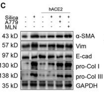
ARG66199 anti-Vimentin antibody [SQab1721] WB image
Western blot: MLE-12 stained with ARG66199 anti-Vimentin antibody [SQab1721].
From Li S et al. Drug Des Devel Ther- (2020), doi: 10.2147/DDDT.S252351, Fig. 4. C.
-
ARG66381 anti-alpha smooth muscle Actin antibody [SQab18108] IHC-P image
Immunohistochemistry: Bovine blood vessels stained with ARG66381 anti-alpha smooth muscle Actin antibody [SQab18108].
From Mari Akiyama et al. Cell Biochem Biophys. (2024), doi: 10.1007/s12013-024-01647-5, Fig. 5.
-
ARG66381 anti-alpha smooth muscle Actin antibody [SQab18108] WB image
Western blot: Rat lung fibroblasts stained with ARG66381 anti-alpha smooth muscle Actin antibody [SQab18108], and ARG59501 anti-TGFBR2 / TGF beta Receptor II antibody.
From Chen Y et al. Preprint- (2020), doi: 10.1016/j.omtn.2019.11.018, Fig. 2. A.
-
ARG66199 anti-Vimentin antibody [SQab1721] WB image
Western blot: 20 µg of HeLa and 293T cell lysates stained with ARG66199 anti-Vimentin antibody [SQab1721] at 1:1000 dilution.
-
ARG66199 anti-Vimentin antibody [SQab1721] ICC/IF image
Immunofluorescence: HeLa cells were fixed with 4% paraformaldehyde for 30 min at RT, permeabilized with 0.1% Triton X-100 for 10 min at RT then blocked with 10% goat serum for 30 min at room temperature. Cells were stained with ARG66199 anti-Vimentin antibody [SQab1721] (green) at 1:25000 and 4°C. DAPI (blue) was used as the nuclear counter stain.
-
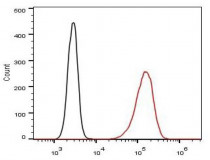
ARG66199 anti-Vimentin antibody [SQab1721] FACS image
Flow Cytometry: HeLa cells were fixed with 4% paraformaldehyde (10 min) and then permeabilized with 0.1% TritonX-100 for 15 min. The cells were then stained with ARG66199 anti-Vimentin antibody [SQab1721] (red) at 1:500 dilution in 1x PBS/1% BSA for 30 min at 4°C, followed by Alexa Fluor® 488 labelled secondary antibody. Unlabelled sample (black) was used as a control.
-
ARG66199 anti-Vimentin antibody [SQab1721] IHC-P image
Immunohistochemistry: Formalin‐fixed and paraffin‐embedded Human colon tissue stained with ARG66199 anti-Vimentin antibody [SQab1721] at 1:1000 dilution.
Antigen retrieval: Heat mediated was performed using Tris/EDTA buffer pH 9.0
-
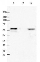
ARG66199 anti-Vimentin antibody [SQab1721] IP image
Immunoprecipitation: 0.4 mg of HeLa cell lysate immunoprecipitated and stained with ARG66199 anti-Vimentin antibody [SQab1721]. 1) IP in HeLa whole cell lysate, 2) Rabbit IgG instead of primary antibody in HeLa whole cell lysate and 3) HeLa whole cell lysate, 10 μg (input).
-
ARG66381 anti-alpha smooth muscle Actin antibody [SQab18108] WB image
Western blot: 30 μg of NRK-49F cells treated with TGF beta 1 (10 ng/ml) for 0~48 hours. Cell lysates were stained with ARG66381 anti-alpha smooth muscle Actin antibody [SQab18108] at 1:2000 dilution, overnight at 4°C.
-
ARG66381 anti-alpha smooth muscle Actin antibody [SQab18108] ICC/IF image
Immunofluorescence: NRK-49F cells treated with TGF beta 1 (10 ng/ml) for 0~48 hours. Cells were fixed with 4% PFA for 15 min at room temperature and permeabilizated by 0.5% Triton X-100. Cells were stained with ARG66381 anti-alpha smooth muscle Actin antibody [SQab18108] at 1:200 dilution, overnight at 4°C.
-
ARG66381 anti-alpha smooth muscle Actin antibody [SQab18108] FACS image
Flow Cytometry: HeLa cells were fixed with 4% paraformaldehyde (10 min) and then permeabilized with 0.1% TritonX-100 for 15 min. The cells were then stained with ARG66381 anti-alpha smooth muscle Actin antibody [SQab18108] (red) at 1:100 dilution in 1x PBS/1% BSA for 30 min at 4°C, followed by Alexa Fluor® 488 labelled secondary antibody. Unlabelled sample (green) was used as a control.
-
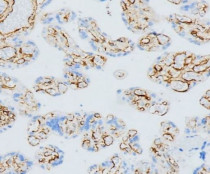
ARG66381 anti-alpha smooth muscle Actin antibody [SQab18108] IHC-P image
Immunohistochemistry: Formalin-fixed and paraffin-embedded placenta stained with ARG66381 anti-alpha smooth muscle Actin antibody [SQab18108] at 1:2000 dilution. Antigen Retrieval: Heat mediated was performed using Tris/EDTA buffer (pH 9.0).
-
ARG66381 anti-alpha smooth muscle Actin antibody [SQab18108] WB image
Western blot: 10 µg of HeLa, HaCat, 293, LnCap, PC-3 and T47D cell lysates stained with ARG66381 anti-alpha smooth muscle Actin antibody [SQab18108] at 1:2000 dilution.
-
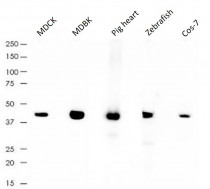
ARG66381 anti-alpha smooth muscle Actin antibody [SQab18108] WB image
Western blot: 10 µg of MDCK, MDBK, Pig heart, Zebrafish and Cos-7 lysates stained with ARG66381 anti-alpha smooth muscle Actin antibody [SQab18108] at 1:2000 dilution.
-
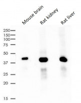
ARG66381 anti-alpha smooth muscle Actin antibody [SQab18108] WB image
Western blot: 10 µg of Mouse brain, Rat kidney and Rat liver lysates stained with ARG66381 anti-alpha smooth muscle Actin antibody [SQab18108] at 1:2000 dilution.
-
ARG66199 anti-Vimentin antibody [SQab1721] WB image
Western blot: 20 µg of COLO205, HCT116, HT29 (Vimentin unexpression cell lines) and SW620 (Vimentin expression cell line). Cell lysates stained with ARG66199 anti-Vimentin antibody [SQab1721] at 1:1000 dilution.
文献引用
