ARG30333
M1 / M2 / TAM Marker Antibody Panel
内含物
| 货号 | 内含物名称 | 宿主克隆性 | 反应 | 应用 | 包装 |
|---|---|---|---|---|---|
| ARG10514 | anti-CD68 antibody [KP1] | Mouse mAb | Hu, Rat | FACS, ICC/IF, IHC-P, WB | 20 μg |
| ARG56509 | anti-iNOS antibody | Rabbit pAb | Hu, Mamm, Ms, Rat | ICC/IF, IHC-P, IHC-Fr, IP, WB | 50 μl |
| ARG55554 | anti-CD206 / MMR antibody [15-2] | Mouse mAb | Hu, Ms | CyTOF®-candidate, FACS, FuncSt, ICC/IF, IHC-Fr, IP, WB | 20 μg |
| ARG66630 | anti-CD163 antibody [SQab19148] | Rabbit mAb | Hu | IHC-P | 20 μl |
概述
| 产品描述 | M1 / M2 / TAM Marker Antibody Panel is an all-in-one solution to make identification of macrophage subtypes easy and economic. This antibody panel comprises M1 macrophage markers CD68 and iNOS antibodies, and M2 macrophage markers CD163 and CD206 antibodies. Besides, all four antibodies are ideal for determining the tumor-associated macrophage (TAM) phenotype. All the antibodies in this panel have excellent IHC staining performance. Also, the different host species make double staining possible. Related news: New antibody panels and duos for Tumor immune microenvironment Anti-SerpinB9 therapy, a new strategy for cancer therapy RIP1 activation and pathogenesis of NASH |
|---|---|
| 靶点名称 | M1 / M2 / TAM Marker |
| 別名 | M1/M2/TAM Marker antibody; CD68 antibody; CD206 / MMR antibody; iNOS antibody; CD163 antibody |
属性
| 存放说明 | For continuous use, store undiluted antibody at 2-8°C for up to a week. For long-term storage, aliquot and store at -20°C or below. Storage in frost free freezers is not recommended. Avoid repeated freeze/thaw cycles. Suggest spin the vial prior to opening. The antibody solution should be gently mixed before use. |
|---|---|
| 注意事项 | For laboratory research only, not for drug, diagnostic or other use. |
生物信息
| 全名 | Antibody Panel for M1/M2/TAM Marker |
|---|---|
| 产品亮点 | Related Product: anti-CD68 antibody; anti-iNOS-antibodies; anti-CD206 / MMR antibody; anti-CD163 antibody; |
检测图片 (17) Click the Picture to Zoom In
-

ARG10514 anti-CD68 antibody [KP1] ICC/IF image
Immunofluorescence: Human colon stained with ARG10514 anti-CD68 antibody [KP1].
From Li Y et al. Research Square (2022), doi: 10.3389/fmicb.2022.926915, Fig. 2C.
-

ARG55554 anti-CD206 / MMR antibody [15-2] ICC/IF image
Immunofluorescence: RAW 264.7 stained with ARG55554 anti-CD206 / MMR antibody [15-2].
From Boda SK et al. Biomater Sci- (2022), doi: 10.1039/d1bm01649k, Fig. 7.
-

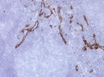
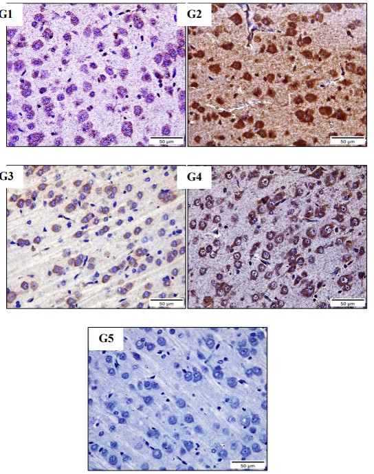
ARG56509 anti-iNOS antibody IHC-P image
Immunohistochemistry: Rat Brain stained with ARG56509 anti-iNOS antibody at 1:100 dilution.
From Abrar Roshdy Abouelkeir et al. European Chemical Bulletin,(2023) doi: 10.31838/ecb/2023.12.1.470, Fig. 6.
-
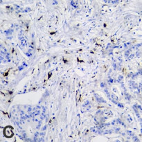
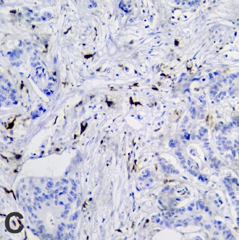
ARG66630 anti-CD163 antibody [SQab19148] IHC-P image
Immunohistochemistry: Human colorectal cancer stained with ARG66630 anti-CD163 antibody [SQab19148].
From Brambilla, Eduardo et al. Doutorado em Ciências da Saúde (2024), URL: https://repositorio.ucs.br/xmlui/handle/11338/13745, Fig. 2. C.
-
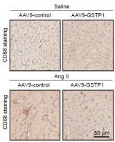
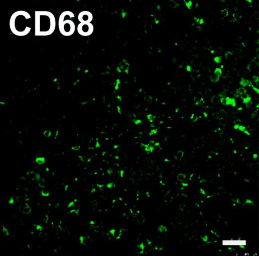
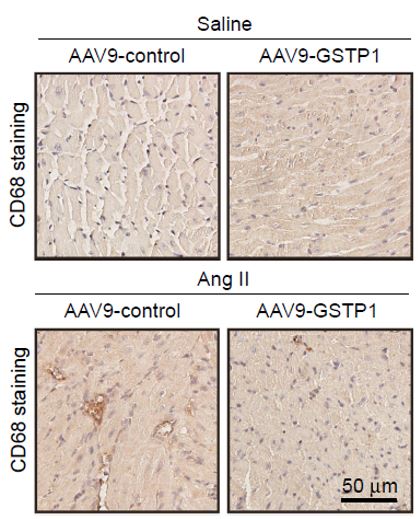
ARG10514 anti-CD68 antibody [KP1] IHC-P image
Immunohistochemistry: Mouse atrial stained with ARG10514 anti-CD68 antibody [KP1].
From Li H et al. Europace (2025), doi: 10.1093/europace/euaf083, Fig. 6E.
-

ARG56509 anti-iNOS antibody ICC/IF image (Customer's Feedback)
Immunofluorescence: RAW264.7 cells were fixed with 4% paraformaldehyde for 15 min at RT, permeabilized with 0.1% Triton X-100 then blocked with 2% albumin for 60 min at RT. Cells were stained with ARG56509 anti-iNOS antibody (green) at 4°C. DAPI (blue) was used as the nuclear counter stain.
-
ARG56509 anti-iNOS antibody WB image
Western blot: Rat Aortic stained with ARG56509 anti-iNOS antibody at 1:1000 dilution.
From Wahid Shah et al. Sci Rep. (2023), doi: 10.1038/s41598-023-43786-4, Fig. 2. C.
-

ARG10514 anti-CD68 antibody [KP1] IHC-P image
Immunohistochemistry: Formalin-fixed and paraffin-embedded Human tonsil stained with ARG10514 anti-CD68 antibody [KP1]. Antigen Retrieval: Boil tissue section in Citrate buffer (pH 6.0).
-
ARG55554 anti-CD206 / MMR antibody [15-2] IHC-Fr image
Immunohistochemistry: Frozen section of Human tonsil tissue stained with ARG55554 anti-CD206 / MMR antibody [15-2] at 1:25 dilution. The antibody stains endothelia of lymph vessels strongly.
-
ARG56509 anti-iNOS antibody IHC-P image
Immunohistochemistry: Paraffin-embedded Human pancreatic ductal adenocarcinoma stained with ARG56509 anti-iNOS antibody.
-

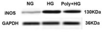
ARG56509 anti-iNOS antibody WB image
Western blot: Raw264.7 cells untreated or treated with LPS. 20 µg of cell lysates stained with ARG56509 anti-iNOS antibody at 1:400 dilution.
-

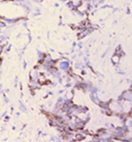
ARG66630 anti-CD163 antibody [SQab19148] IHC-P image
Immunohistochemistry: Formalin/PFA-fixed and paraffin-embedded Human liver tissue stained with ARG66630 anti-CD163 antibody [SQab19148]. Antigen Retrieval: Heat mediation was performed in Tris/EDTA buffer (pH 9.0).
-
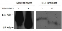
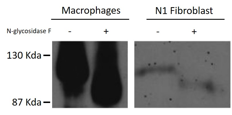
ARG10514 anti-CD68 antibody [KP1] WB image
Western blot: Human Macrophages and N1 Fibroblast untreated or treated with N-glycosidase F. The blots were stained with ARG10514 anti-CD68 antibody [KP1].
-
ARG55554 anti-CD206 / MMR antibody [15-2] FACS image
Flow Cytometry: Stimulated (GM-CSF + IL-4) human peripheral blood mononuclear cells stained with ARG55554 anti-CD206 / MMR antibody [15-2] at 9 µg/ml dilution, followed by PE-conjugated Goat anti-Mouse antibody.
-

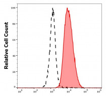
ARG10514 anti-CD68 antibody [KP1] FACS image
Flow Cytometry: THP1 cells prefixed with 4% PFA and then permeabilised with 0.25% saponin. Cells were stained with ARG10514 anti-CD68 antibody [KP1] (white area) or isotype control antibody (gray area).
-

ARG10514 anti-CD68 antibody [KP1] IHC-P image
Immunohistochemistry: Formalin-fixed and paraffin-embedded Human breast carcinoma stained with ARG10514 anti-CD68 antibody [KP1].
-
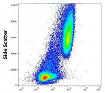
ARG55554 anti-CD206 / MMR antibody [15-2] FACS image
Flow Cytometry: Separation of human CD206 positive dendritic cells differentiated upon monocyte stimulation (GM-CSF + IL-4) (red-filled) from non-stimulated lymphocytes (black-dashed). Human stimulated (GM-CSF + IL-4) peripheral blood mononuclear cells stained with ARG55554 anti-CD206 / MMR antibody [15-2] at 9 µg/ml dilution, followed by PE-conjugated Goat anti-Mouse antibody.


文献引用
Hierarchical Assembly and Dynamic Reinforced Woven Biotextiles for Anisotropic and Viscoelastic Tendon Tissue Regeneration
ARG10514; IHC-P / Mouse


Exploring macrophage polarization as a prognostic indicator for colorectal cancer
ARG10514; IHC-P / Human
Necroptosis of hippocampal neurons in paclitaxel chemotherapy-induced cognitive impairment mediates microglial activation via TLR4/MyD88 signaling pathway
ARG56509; IHC-Fr / Mouse
GSTP1 inhibits angiotensin II-induced atrial fibrillation by regulating ferroptosis
ARG10514; IHC-P / Mouse
Melatonin receptor 1A/B knockout accelerates atrial aging in mice
ARG56509; IHC-P / Mouse
Facile and rapid fabrication of a novel 3D-printable, visible light-crosslinkable and bioactive polythiourethane for large-to-massive rotator cuff tendon repair
ARG10514: IHC-P / Mouse
Melatonin alleviates aging-related heart failure through melatonin receptor 1A/B knockout in mice
ARG56509: IHC-P / Mouse
Banxia-Houpu decoction inhibits iron overload and chronic intermittent hypoxia-induced neuroinflammation in mice
ARG56509: WB, IHC-P / Mouse
Polarização dos macrófagos e sua associação com fatores prognósticos em casos de neoplasias colorretais, considerando a presença e ausência de mutações nas metaloproteinases
ARG66630: IHC-P / Human
Mac-1 deficiency ameliorates pressure overloaded heart failure through inhibiting macrophage polarization and activation
ARG10514: IHC-Fr / Mouse
Cardamonin intervenes in myocardial hypertrophy progression by regulating Usp18
ARG10514: IHC-P / Mouse
Identification and experimental verification of immune-related hub genes in intervertebral disc degeneration
ARG10514: ICC/IF / Human
Effect of macrophage-to-myofibroblast transition on silicosis
ARG56509: IHC-P,WB / Rat
Aquaporin-9 Aggravates Lipopolysaccharides Induced Acute Lung Injury Via Facilitating M1-Like Macrophage Polarization
ARG10514: IHC-P / Mouse
Chronic intermittent hypobaric hypoxia alleviates early-stage posttraumatic osteoarthritis via NF-κB/Nrf2 pathway in mice
ARG56509; WB / Mouse
Polydatin improves vascular endothelial function by maintaining mitochondrial homeostasis under high glucose conditions
ARG56509: WB / Rat
Anti-Inflammatory Actions of G-Protein-Coupled Estrogen Receptor 1 (GPER) and Brain-Derived Estrogen Following Cerebral Ischemia in Ovariectomized Rats
ARG56509: WB, IHC-Fr / Rat
Berberine ameliorates mesenteric vascular dysfunction by modulating perivascular adipose tissue in diet-induced obese in rats
ARG56509: IHC-P / Rat
Paclitaxel induces cognitive impairment via necroptosis, decreased synaptic plasticity and M1 polarisation of microglia
ARG56509: IHC-Fr / Mouse
Dual keratinocyte-attachment and anti-inflammatory coatings for soft tissue sealing around transmucosal oral implants
ARG56509: ICC/IF / Mouse
The Proinflammatory Role of Guanylate-Binding Protein 5 in Inflammatory Bowel Diseases
ARG10514: ICC/IF / Mouse
Complement-mediated M2/M1 macrophage polarization may be involved in crescent formation in lupus nephritis
ARG10514: IHC-P / Human
Glycolytic Reprogramming in Silica-Induced Lung Macrophages and Silicosis Reversed by Ac-SDKP Treatment
ARG56509: WB / Rat
Btk knockout attenuates the liver inflammation in STZ-induced diabetic mice by suppressing NLRP3 inflammasome activation.
ARG10514: IHC-P / Mouse
Enhanced Bruton's tyrosine kinase activity in the kidney of patients with IgA nephropathy.
ARG10514: IHC-P / Human
TAB1 regulates glycolysis and activation of macrophages in diabetic nephropathy.
ARG10514: IHC-P / Mouse
Baihe Wuyao decoction ameliorates CCl 4-induced chronic liver injury and liver fibrosis in mice through blocking TGF-β1/Smad2/3 signaling, anti-inflammation and anti-oxidation effects.
ARG56509: WB / Mouse
A free-standing multilayer film as a novel delivery carrier of platelet lysates for potential wound-dressing applications.
ARG56509: IHC-P / Rat
Exosomes from high glucose-treated macrophages activate glomerular mesangial cells via TGF-β1/Smad3 pathway in vivo and in vitro.
ARG10514: IHC-P / Mouse
HYCO-3, a dual CO-releaser/Nrf2 activator, reduces tissue inflammation in mice challenged with lipopolysaccharide.
ARG56509: IHC-P / Mouse
Distribution and proportion of M1/M2 macrophages in periodontal tissues in rats with and without periodontitis
ARG56509: IHC-P / Rat


