ARG30336
Human MDSC Marker Antibody Duo
Myeloid-derived suppressor cells marker; MDSCs marker; Tumor immune microenvironment; tumor-associated myeloid cells
内含物
| 货号 | 内含物名称 | 宿主克隆性 | 反应 | 应用 | 包装 |
|---|---|---|---|---|---|
| ARG22000 | anti-CD11b antibody [M1/70] | Rat mAb | Bb, Hu, Ms, R. Mk | BL, FACS, ICC/IF, IHC-Fr, IP | 50 μg |
| ARG65560 | anti-CD33 antibody [WM53] | Mouse mAb | Hu, NHuPrm | CyTOF®-candidate, FACS, FuncSt, ICC/IF, IHC-Fr, IP, WB | 50 μg |
概述
| 产品描述 | Human MDSC Marker Antibody Duo includes antibodies to MDSC marker, CD11b and CD33, for Myeloid-derived suppressor cells (MDSCs) study. Myeloid-derived suppressor cells (MDSCs) are a heterogeneous population of immune cells from the myeloid lineage. MDSCs are well-known key negative regulators of the immune response in tumor microenvironment. Accumulated evidences show that MDSCs may serve as a therapeutic target for preventing tumor progression.In humam, MDSCs are defined as cells expressing CD33 and CD11b. arigo’s Human MDSC Marker Antibody Duo comprises CD33 and CD11b antibodies. It is the best solution to identify the MDSCs in human tumor tissues. Related news: New antibody panels and duos for Tumor immune microenvironment Anti-SerpinB9 therapy, a new strategy for cancer therapy |
|---|---|
| 靶点名称 | Human MDSC marker |
| 別名 | Human MDSC Marker antibody; Myeloid-derived suppressor cells marker antibody; MDSCs marker antibody; CD11b antibody; CD33 antibody |
属性
| 存放说明 | For continuous use, store undiluted antibody at 2-8°C for up to a week. For long-term storage, aliquot and store at -20°C or below. Storage in frost free freezers is not recommended. Avoid repeated freeze/thaw cycles. Suggest spin the vial prior to opening. The antibody solution should be gently mixed before use. |
|---|---|
| 注意事项 | For laboratory research only, not for drug, diagnostic or other use. |
生物信息
| 全名 | Human Myeloid-derived suppressor cell (MDSC) marker Antibody Duo |
|---|---|
| 产品亮点 | Related products: anti-CD11b antibody; anti-CD33 antibody; MDSC Duos / Panels; |
| 研究领域 | Myeloid-derived suppressor cells marker; MDSCs marker; Tumor immune microenvironment; tumor-associated myeloid cells |
检测图片 (7) Click the Picture to Zoom In
-
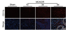
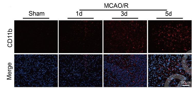
ARG22000 anti-CD11b antibody [M1/70] IHC-Fr image
Immunohistochemistry: Frozen Mouse brain stained with ARG22000 anti-CD11b antibody [M1/70].
From Gui-Nan Jiang et al. SSRN (2025), doi: 10.2139/ssrn.5219692, Fig. 1C.
-
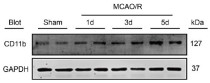
ARG22000 anti-CD11b antibody [M1/70] WB image
Western blot: Mouse brain stained with ARG22000 anti-CD11b antibody [M1/70].
From Gui-Nan Jiang et al. SSRN (2025), doi: 10.2139/ssrn.5219692, Fig. 1B.
-
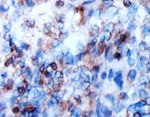
ARG65560 anti-CD33 antibody [WM53] IHC-Fr image
Immunohistochemistry: Frozen section of Human colon tissue stained with ARG65560 anti-CD33 antibody [WM53].
-
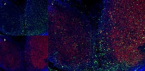
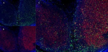
ARG22000 anti-CD11b antibody [M1/70] IHC-Fr image
Immunohistochemistry: Cryosection of Mouse lymph node stained with ARG22000 anti-CD11b antibody [M1/70] (green, A) and anti-Mouse CD8 antibody (red, B). Merged image in C.
-
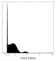
ARG65560 anti-CD33 antibody [WM53] CyTOF image
CyTOF: Human peripheral blood stained with ARG65560 anti-CD33 antibody [WM53] (149Sm). Singlet cells were gated for data analysis.
-
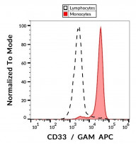
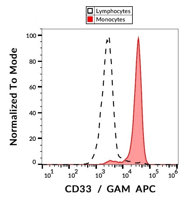
ARG65560 anti-CD33 antibody [WM53] FACS image
Flow Cytometry: Separation of Human CD33 positive Monocytes (red) from Human CD33 negative Lymphocytes (black-dashed). Human peripheral blood stained with ARG65560 anti-CD33 antibody [WM53], followed by incubation with APC labelled Goat anti-Mouse secondary antibody.
-
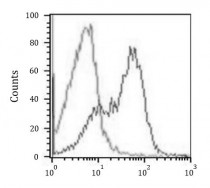
ARG65560 anti-CD33 antibody [WM53] FACS image
Flow Cytometry: PBMC stained with ARG65560 anti-CD33 antibody [WM53] at 0.5 µg/10^6 cells (right histogram) or isotype control (left histogram), followed by incubation with PE labelled secondary antibody.


文献引用
Integrin CD11b alleviates cerebral ischemia/reperfusion injury through a mechanism involving microglia/macrophage polarization
ARG22000; WB, IHC-Fr / Mouse


Acupuncture promotes neurological recovery and regulates lymphatic function after acute inflammatory nerve root injury
ARG22000: IHC-Fr / Human
Mac-1 deficiency ameliorates pressure overloaded heart failure through inhibiting macrophage polarization and activation
ARG22000: IHC-Fr, WB / Mouse
CD11b mediates hypertensive cardiac remodeling by regulating macrophage infiltration and polarization
Mouse / ARG22000: IHC-P, WB
Integrin CD11b Contributes to Hypertension and Vascular Dysfunction Through Mediating Macrophage Adhesion and Migration
ARG22000:WB / Mouse


