ARG65560
anti-CD33 antibody [WM53]
anti-CD33 antibody [WM53] for CyTOF®-candidate,Flow cytometry,Functional study,ICC/IF,IHC-Frozen sections,Immunoprecipitation,Western blot and Human,Primates
Developmental Biology antibody; Immune System antibody; Human MDSC Marker antibody; Myeloid-derived suppressor cell antibody
概述
| 产品描述 | Mouse Monoclonal antibody [WM53] recognizes CD33 |
|---|---|
| 反应物种 | Hu, NHuPrm |
| 应用 | CyTOF®-candidate, FACS, FuncSt, ICC/IF, IHC-Fr, IP, WB |
| 特异性 | The clone WM53 reacts with CD33, a 67 kDa type I transmembrane glycoprotein (immunoglobulin superfamily) expressed on myeloid progenitors, monocytes, granulocytes, dendritic cells and mast cells; it is absent on platelets, lymphocytes, erythrocytes and hematopoietic stem cells. HLDA IV; WS Code M-505 |
| 宿主 | Mouse |
| 克隆 | Monoclonal |
| 克隆号 | WM53 |
| 同位型 | IgG1 |
| 靶点名称 | CD33 |
| 抗原物种 | Human |
| 抗原 | Human AML cells |
| 偶联标记 | Un-conjugated |
| 別名 | p67; Sialic acid-binding Ig-like lectin 3; SIGLEC-3; CD antigen CD33; gp67; Siglec-3; Myeloid cell surface antigen CD33; SIGLEC3 |
应用说明
| 应用建议 |
|
||||||||||||||||
|---|---|---|---|---|---|---|---|---|---|---|---|---|---|---|---|---|---|
| 应用说明 | IHC-Fr: Acetone fixation. Functional application: Induction of cytokine production. * The dilutions indicate recommended starting dilutions and the optimal dilutions or concentrations should be determined by the scientist. |
属性
| 形式 | Liquid |
|---|---|
| 纯化 | Purified from cell culture supernatant by protein-A affinity chromatography. |
| 纯度 | > 95% (by SDS-PAGE) |
| 缓冲液 | PBS (pH 7.4) and 15 mM Sodium azide |
| 抗菌剂 | 15 mM Sodium azide |
| 浓度 | 0.1 mg/ml |
| 存放说明 | For continuous use, store undiluted antibody at 2-8°C for up to a week. For long-term storage, aliquot and store at -20°C or below. Storage in frost free freezers is not recommended. Avoid repeated freeze/thaw cycles. Suggest spin the vial prior to opening. The antibody solution should be gently mixed before use. |
| 注意事项 | For laboratory research only, not for drug, diagnostic or other use. |
生物信息
| 数据库连接 | |
|---|---|
| 基因名称 | CD33 |
| 全名 | CD33 molecule |
| 背景介绍 | CD33 is a transmembrane protein of the sialic acid-binding immunoglobulin-like lectin (Siglec) family. It belongs to the immunoreceptor tyrosine-based inhibitory motif (ITIM)-containing molecules able of recruiting protein tyrosine phosphatases SHP-1 and SHP-2 to signal assemblies; these ITIMs are also used for ubiquitin-mediated removal of the receptor from the cell surface. CD33 is expressed on cells of myelomonocytic lineage, binds sialic acid residues in N- and O-glycans on cell surfaces, and is a therapeutic target for acute myeloid leukemia. |
| 生物功能 | CD33: Sialic-acid-binding immunoglobulin-like lectin (Siglec) that plays a role in mediating cell-cell interactions and in maintaining immune cells in a resting state (PubMed:10611343, PubMed:15597323, PubMed:11320212). Preferentially recognizes and binds alpha-2,3- and more avidly alpha-2,6-linked sialic acid-bearing glycans (PubMed:7718872). Upon engagement of ligands such as C1q or syalylated glycoproteins, two immunoreceptor tyrosine-based inhibitory motifs (ITIMs) located in CD33 cytoplasmic tail are phosphorylated by Src-like kinases such as LCK (PubMed:28325905, PubMed:10887109). These phosphorylations provide docking sites for the recruitment and activation of protein-tyrosine phosphatases PTPN6/SHP-1 and PTPN11/SHP-2 (PubMed:10556798, PubMed:10206955, PubMed:10887109). In turn, these phosphatases regulate downstream pathways through dephosphorylation of signaling molecules (PubMed:10206955, PubMed:10887109). One of the repressive effect of CD33 on monocyte activation requires phosphoinositide 3-kinase/PI3K (PubMed:15597323). [UniProt] |
| 产品亮点 | Related Antibody Duos and Panels: ARG30336 Human MDSC Marker Antibody Duo Related products: CD33 antibodies; CD33 ELISA Kits; CD33 Duos / Panels; Anti-Mouse IgG secondary antibodies; Related news: CyTOF-candidate Antibodies New antibody panels and duos for Tumor immune microenvironment Anti-SerpinB9 therapy, a new strategy for cancer therapy |
| 研究领域 | Developmental Biology antibody; Immune System antibody; Human MDSC Marker antibody; Myeloid-derived suppressor cell antibody |
| 预测分子量 | 40 kDa |
| 翻译后修饰 | Phosphorylation of Tyr-340 is involved in binding to PTPN6 and PTPN11. Phosphorylation of Tyr-358 is involved in binding to PTPN6. |
检测图片 (4) Click the Picture to Zoom In
-
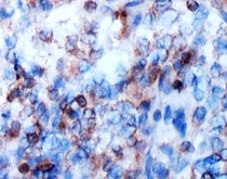
ARG65560 anti-CD33 antibody [WM53] IHC-Fr image
Immunohistochemistry: Frozen section of Human colon tissue stained with ARG65560 anti-CD33 antibody [WM53].
-
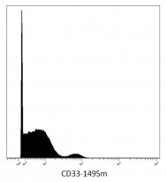
ARG65560 anti-CD33 antibody [WM53] CyTOF image
CyTOF: Human peripheral blood stained with ARG65560 anti-CD33 antibody [WM53] (149Sm). Singlet cells were gated for data analysis.
-
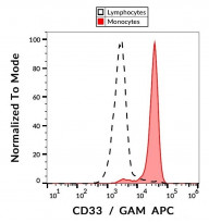
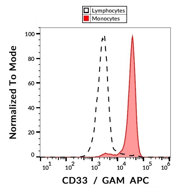
ARG65560 anti-CD33 antibody [WM53] FACS image
Flow Cytometry: Separation of Human CD33 positive Monocytes (red) from Human CD33 negative Lymphocytes (black-dashed). Human peripheral blood stained with ARG65560 anti-CD33 antibody [WM53], followed by incubation with APC labelled Goat anti-Mouse secondary antibody.
-
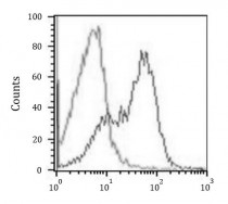
ARG65560 anti-CD33 antibody [WM53] FACS image
Flow Cytometry: PBMC stained with ARG65560 anti-CD33 antibody [WM53] at 0.5 µg/10^6 cells (right histogram) or isotype control (left histogram), followed by incubation with PE labelled secondary antibody.


克隆号文献
Highly multiparametric analysis by mass cytometry.
FACS / Human


A study of CD33 (SIGLEC-3) antigen expression and function on activated human T and NK cells: two isoforms of CD33 are generated by alternative splicing.
Mobilization of bone marrow-derived stem cells after myocardial infarction and left ventricular function.
Macrophages expressing triggering receptor expressed on myeloid cells-1 are underrepresented in the human intestine.
Expression of the myeloid-associated marker CD33 is not an exclusive factor for leukemic plasmacytoid dendritic cells.


