ARG30151
Glucose uptake: Insulin Receptor Dependent Pathway Antibody Panel (GLUT4, AKT pS473, IRS1 pS636)
Cancer antibody; Cell Biology and Cellular Response antibody; Cell Death antibody; Controls and Markers antibody; Developmental Biology antibody; Gene Regulation antibody; Immune System antibody; Metabolism antibody; Neuroscience antibody; Signaling Transduction antibody
内含物
| 货号 | 内含物名称 | 宿主克隆性 | 反应 | 应用 | 包装 |
|---|---|---|---|---|---|
| ARG51720 | anti-IRS1 phospho (Ser636) antibody | Rabbit pAb | Hu, Ms, Rat | IHC-P, WB | 50 μl |
| ARG51558 | anti-Akt phospho (Ser473) antibody | Rabbit pAb | Hu, Ms, Rat | ICC/IF, IHC-P, WB | 50 μl |
| ARG65292 | anti-GLUT4 antibody | Goat pAb | Hu, Ms, Rat | IHC-P, WB | 50 μg |
| ARG65351 | Goat anti-Rabbit IgG antibody (HRP) | Goat pAb | Rb | ELISA, IHC-P, WB | 50 μl |
| ARG65352 | Donkey anti-Goat IgG antibody (HRP) | Donkey pAb | Goat | ELISA, IHC-P, WB | 50 μl |
概述
| 产品描述 | Glucose is the primary energy source for most cells of the body. The study of glucose metabolism is central to cell proliferation, growth, survival, and most recently, tumor progression. Insulin stimulates glucose uptake from blood into skeletal muscle and adipose tissue through a signaling cascade mediated by the insulin receptor (IR). Insulin binding to the IR results in activation of the insulin receptor substrate (IRS) protein and subsequent signaling to the PI3K/Akt pathways, resulting in translocation of Glut4 vesicles, glucose uptake, cell proliferation, and survival. Otherwise, glucose uptake also can be stimulated by adrenoceptor. Upon ligand binding, b3-adrenoceptors result in increased levels of intracellular cAMP that lead to increased de novo synthesis of GLUT1 and mTORC2 phosphorylation. The newly produced GLUT1 is then translocated to the plasma membrane and helps the glucose uptake from blood. These two antibody PANELs can be used to identify the glucose uptake through insulin or adrenoceptors dependent pathway. Mihaylova MM, Shaw RJ (2011) Nat. Cell Biol. 13(9), 1016–23. Zemva J, Schubert M (2011) Curr Diabetes Rev 7(5), 356–66. Olsen, J. M. et al (2014) J. Cell Biol.207(3), 365-374. Related news: Quantifying IR-associated Adipokines with Multiplex ELISA kit; |
|---|---|
| 靶点名称 | Glucose uptake: Insulin Receptor Dependent Pathway |
| 別名 | Glucose uptake: Insulin Receptor Dependent Pathway antibody; Akt phospho (Ser473) antibody; IRS1 phospho (Ser636) antibody; GLUT4 antibody |
属性
| 存放说明 | For continuous use, store undiluted antibody at 2-8°C for up to a week. For long-term storage, aliquot and store at -20°C or below. Storage in frost free freezers is not recommended. Avoid repeated freeze/thaw cycles. Suggest spin the vial prior to opening. The antibody solution should be gently mixed before use. |
|---|---|
| 注意事项 | For laboratory research only, not for drug, diagnostic or other use. |
生物信息
| 全名 | Antibody Panel for Glucose uptake: Insulin Receptor Dependent Pathway (GLUT4, AKT pS473, IRS1 pS636) |
|---|---|
| 产品亮点 | Related Product: anti-GLUT4 antibody; |
| 研究领域 | Cancer antibody; Cell Biology and Cellular Response antibody; Cell Death antibody; Controls and Markers antibody; Developmental Biology antibody; Gene Regulation antibody; Immune System antibody; Metabolism antibody; Neuroscience antibody; Signaling Transduction antibody |
检测图片 (18) Click the Picture to Zoom In
-
ARG51558 anti-Akt phospho (Ser473) antibody IHC-P image
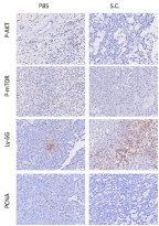
Immunohistochemistry: Mouse xenograft tumor stained with ARG22104 anti-Ly6G + Ly6C antibody [RB6-8C5] , ARG40666 anti-mTOR phospho (Ser2448) antibody , ARG51558 anti-Akt phospho (Ser473) antibodyand ARG62605 anti-PCNA antibody [PC10] .
From Wu LH et al. J Cancer (2022), doi: 10.7150/jca.75163, Fig. 5. D.
-
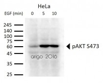
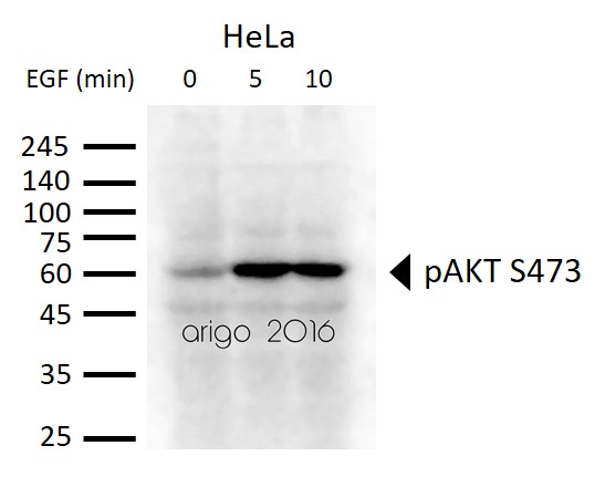
ARG51558 anti-Akt phospho (Ser473) antibody WB image
Western blot: 30 µg of HeLa cells untreated or treated with EGF. Lysates stained with ARG51558 anti-Akt phospho (Ser473) antibody at 1:500 dilution.
-
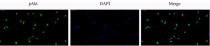
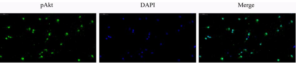
ARG51558 anti-Akt phospho (Ser473) antibody ICC-IF image
Immunofluorescence: Rat neurons stained with ARG51558 anti-Akt phospho (Ser473) antibody.
From Gao Z et al. Neuroreport- (2021), doi: 10.1097/WNR.0000000000001593, Fig. 4.
-
_210_205.jpg)
ARG65352 Donkey anti-Goat IgG antibody (HRP) WB image
Western blot: Endometrial Stromal cells stained with ARG63922 anti-EP4 prostaglandin Receptor antibody at 1:1000 dilution and ARG65352 Donkey anti-Goat IgG antibody (HRP).
From Qingqing Huang et al. Reprod Med Biol. (2021), doi: 10.1002/rmb2.12423, Fig. 3.C.
-
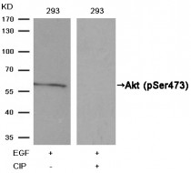
ARG51558 anti-Akt phospho (Ser473) antibody WB image
Western Blot: extracts from 293 cells, treatedwith EGF or calf intestinal phosphatase (CIP), stained with anti-Akt (phospho Ser473) antibody ARG51558.
-
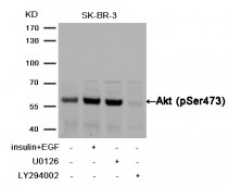
ARG51558 anti-Akt phospho (Ser473) antibody WB image
Western Blot: extracts from SK-BR-3 cells,treated with insulin and EGF, and pretreated with U0126and LY294002 cells stained with anti-Akt (phospho Ser473) antibody ARG51558.
-
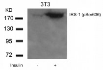
ARG51720 anti-IRS-1 phospho (Ser636) antibody WB image
Western Blot: extracts from 3T3 cells untreated or treated with Insulin stained with anti-IRS-1 (phospho Ser636) antibody ARG51720.
-
ARG65292 anti-SLC2A4 / GLUT4 antibody WB image
Western Blot: Mouse Heart lysate (35 µg protein in RIPA buffer) stained with ARG65292 anti-SLC2A4 / GLUT4 antibody at 1 µg/ml dilution.
-
ARG51558 anti-Akt phospho (Ser473) antibody WB image
Western blot: HepG2 stained with ARG51558 anti-Akt phospho (Ser473) antibody and ARG51712 anti-mTOR phospho (Ser2448) antibody.
From Lu J et al. Toxicology- (2021), doi: 10.1016/j.tox.2021.152716, Fig. 2. A.
-
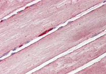
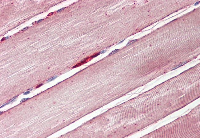
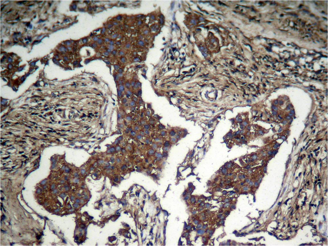
ARG65292 anti-GLUT4 antibody IHC-P image
Immunohistochemistry: Paraffin-embedded Human skeletal muscle tissue. Antigen Retrieval: Steam tissue section in Citrate buffer (pH 6.0). The tissue section was stained with ARG65292 anti-GLUT4 antibody at 3.75 µg/ml dilution followed by AP-staining.
-
ARG51558 anti-Akt phospho (Ser473) antibody WB image
Western blot: Rat neurons stained with ARG51558 anti-Akt phospho (Ser473) antibody.
From Gao Z et al. Neuroreport- (2021), doi: 10.1097/WNR.0000000000001593, Fig. 3. A.
-

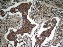
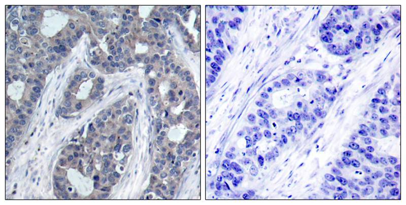
ARG51558 anti-Akt phospho (Ser473) antibody IHC-P image
Immunohistochemistry: paraffin-embedded human breast carcinoma tissue stained with anti-Akt (phospho Ser473) antibody ARG51558 (left) or the same antibody preincubated with blocking peptide (right).
-
ARG51558 anti-Akt phospho (Ser473) antibody IHC-P image
Immunohistochemistry: paraffinembeddedhuman Lung carcinoma tissue stained with anti-Akt (phospho Ser473) antibody ARG51558.
-

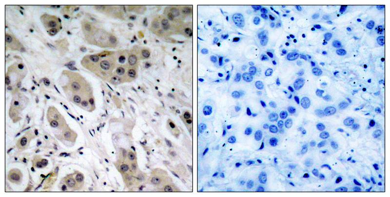
ARG51720 anti-IRS-1 phospho (Ser636) antibody IHC-P image
Immunohistochemistry: paraffin-embedded human breast carcinoma tissue stained with anti-IRS-1 (phospho Ser636) antibody ARG51720 (left) or the same antibody preincubated with blocking peptide (right).
-
ARG51558 anti-Akt phospho (Ser473) antibody WB image
Western blot: PC12 stained with ARG51558 anti-Akt phospho (Ser473) antibody.
From Liu Y et al. Pharmaceuticals (Basel). (2024), doi: 10.3390/ph17070972, Fig. 4. A.
-
ARG51558 anti-Akt phospho (Ser473) antibody WB image
Western blot: Mouse heart stained with ARG51558 anti-Akt phospho (Ser473) antibody and ARG56418 anti-Akt (pan) antibody.
From Lin QY et al. Am J Physiol Heart Circ Physiol- (2019), doi: 10.1152/ajpheart.00566.2019, Fig. 3. D.
-

ARG65351 Goat anti-Rabbit IgG antibody (HRP) WB image
Western blot: Gastric cancer cells stained with ARG66247 anti-Bax antibody, ARG55188 anti-Bcl 2 antibody, ARG57512 anti-Caspase 3 (cleaved) antibody and ARG62346 anti-beta Actin antibody [BA3R].
Secondary Antibody stained with ARG65351 Goat anti-Rabbit IgG antibody (HRP).From Limin Zhang et al. Heliyon (2024), doi: 10.1016/j.heliyon.2024.e30803, Fig. 4. C.
-

ARG65351 Goat anti-Rabbit IgG antibody (HRP) WB image
Western blot: Rat placental stained with ARG57589 anti-MTNR1A antibody at 1:1000 dilution, ARG65351 Goat anti-Rabbit IgG antibody (HRP) at 1:5000 dilution.
From Jinzhi Li et al. J Reprod Immunol. (2023), doi: 10.1016/j.jri.2023.104166, Fig. 2.B.

.jpg)


文献引用
Soybean Trypsin Inhibitor Possesses Potency Against SARS-CoV-2 Infection by Blocking the Host Cell Surface Receptors ACE2, TMPRSS2, and CD147
ARG65351; WB /


Progressively diminished estrogen signaling concordant with increased fibrosis in ectopic endometrium
ARG65351; WB /
The Therapeutic Potential of Exosomes vs. Matrix-Bound Nanovesicles from Human Umbilical Cord Mesenchymal Stromal Cells in Osteoarthritis Treatment
ARG65351; WB /
Environmental acidification drives inter-organ energy mobilization to enhance reproductive performance in medaka (Oryzias latipes)
ARG65351; WB /
Sika Deer Velvet Antler Peptide Exerts Neuroprotective Effect in a Parkinson's Disease Model via Regulating Oxidative Damage and Gut Microbiota
ARG51558: WB / Mouse
Downregulation of AKT/mTOR signaling pathway for Salmonella-mediated autophagy in human anaplastic thyroid cancer
ARG51558: IHC-P / Mouse
Mild hypothermia protects rat cortical neurons against oxygen-glucose deprivation/reoxygenation injury via the PI3K/Akt pathway.
ARG51558: WB, ICC/IF / Rat
Dopamine D2 receptor regulates cortical synaptic pruning in rodents
ARG51558: WB / Rat
1,3-dichloro-2-propanol induced lipid accumulation by blocking autophagy flux in HepG2 cells.
ARG51558: WB / Human
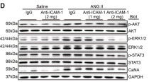
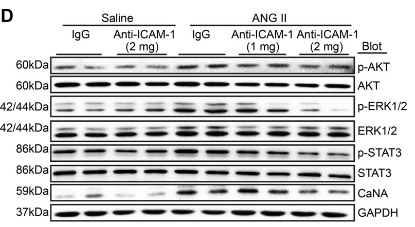
Pharmacological blockage of ICAM-1 improves angiotensin II-induced cardiac remodeling by inhibiting adhesion of LFA-1+ monocytes.
ARG51558: WB / Mouse
Protective effects of dioscin against fructose-induced renal damage via adjusting Sirt3-mediatedoxidative stress, fibrosis, lipid metabolism and inflammation.
ARG51558: WB / Rat
电针对控制性超排卵小鼠子宫内膜磷酸化胰岛素样生长因子-1受体表达及其信号转导通路的调控
ARG51558: WB / Mouse


