ARG30259
Loading Controls for Cytoplasmic / Nuclear Fractions Antibody Panel
Cancer antibody; Controls and Markers antibody; Gene Regulation antibody; Immune System antibody; Metabolism antibody; Neuroscience antibody; Signaling Transduction antibody
内含物
| 货号 | 内含物名称 | 宿主克隆性 | 反应 | 应用 | 包装 |
|---|---|---|---|---|---|
| ARG54003 | anti-COX4 antibody | Mouse mAb | Hu, Ms, Rat, Goat, Hm, Mk | FACS, ICC/IF, IHC-P, IP, WB | 25 μl |
| ARG65681 | anti-Histone H3 antibody | Mouse mAb | Hu, Ms, Rat | ICC/IF, IHC-P, IP, WB | 25 μg |
| ARG10112 | anti-GAPDH antibody [6C5] | Mouse mAb | AGMK, Bb, Cat, Chk, Dog, Fsh, Hm, Hu, Mk, Ms, Pig, Rb, Rat, Xenopus laevis, Zfsh | ELISA, ICC/IF, IHC-Fr, WB | 25 μg |
| ARG65350 | Goat anti-Mouse IgG antibody (HRP) | Goat pAb | Ms | ELISA, IHC-P, WB | 50 μl |
概述
| 靶点名称 | Loading Controls for Cytoplasmic / Nuclear Fractions |
|---|---|
| 別名 | Loading Controls for Cytoplasmic / Nuclear Fractions antibody; GAPDH antibody; COX4 antibody; Histone H3 antibody |
属性
| 存放说明 | For continuous use, store undiluted antibody at 2-8°C for up to a week. For long-term storage, aliquot and store at -20°C or below. Storage in frost free freezers is not recommended. Avoid repeated freeze/thaw cycles. Suggest spin the vial prior to opening. The antibody solution should be gently mixed before use. |
|---|---|
| 注意事项 | For laboratory research only, not for drug, diagnostic or other use. |
生物信息
| 全名 | Antibody Panel for Loading Controls for Cytoplasmic / Nuclear Fractions |
|---|---|
| 产品亮点 | Related Product: anti-Histone H3 antibody; anti-GAPDH antibody; |
| 研究领域 | Cancer antibody; Controls and Markers antibody; Gene Regulation antibody; Immune System antibody; Metabolism antibody; Neuroscience antibody; Signaling Transduction antibody |
检测图片 (27) Click the Picture to Zoom In
-
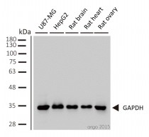
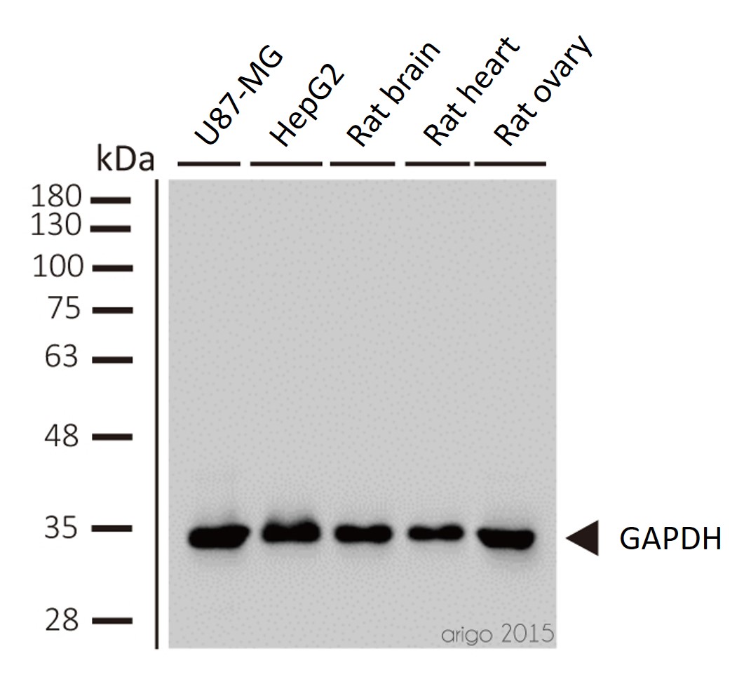
ARG10112 anti-GAPDH antibody [6C5] WB image
Western blot: 1) U87-MG 2) HepG2 3) rat brain 4) rat heart 5) rat ovary stained with ARG10112 anti-GAPDH antibody [6C5] at 1:2000 dilution.
-

ARG65681 anti-Histone H3 antibody IP image
Immunoprecipitation: 1) HeLa cell lysate stained with ARG65681 anti-Histone H3 antibody and 2) IP product immunoprecipitated by ARG65681 anti-Histone H3 antibody at 1:200 dilution.
-

ARG10112 anti-GAPDH antibody [6C5] WB image
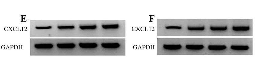
Western blot: HK2 stained with ARG66421 anti-CXCL12 / SDF1 antibody [SQab18123] and ARG10112 anti-GAPDH antibody [6C5] and secondary antibody used ARG65351 Goat anti-Rabbit IgG antibody (HRP).
From Zhou Y et al. Central European Journal of Immunology (2022), doi: 10.5114/ceji.2022.115628, Fig. 2. E.
-
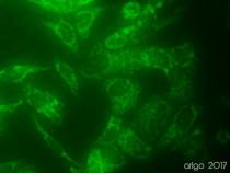
ARG54003 anti-COX4 antibody ICC/IF image
Immunofluorescence: 100% Methanol fixed (RT, 10 min) HeLa cells stained with ARG54003 anti-COX4 antibody (green) at 1:150 dilution.
Secondary antibody: ARG55393 Goat anti-Mouse IgG (H+L) antibody (FITC)
-

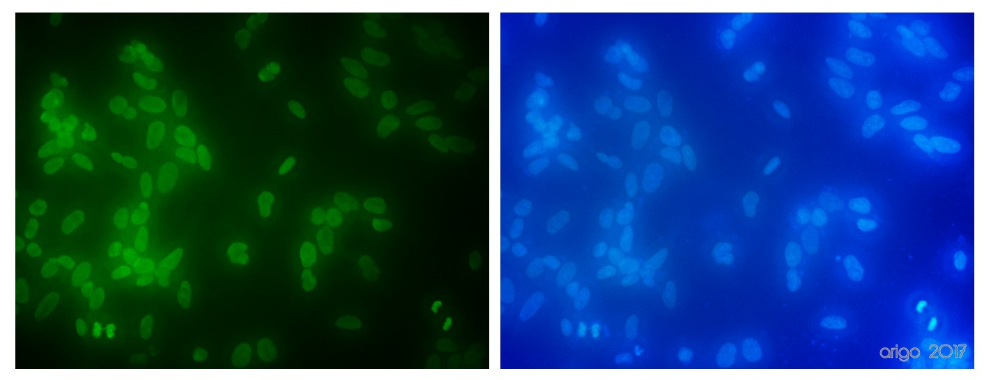
ARG65681 anti-Histone H3 antibody ICC/IF image
Immunofluorescence: 100% Methanol fixed (RT, 10 min) HeLa cells stained with ARG65681 anti-Histone H3 antibody at 1:100 dilution. Left: primary antibody (green). Right: Merge (primary antibody and DAPI).
Secondary antibody: ARG55393 Goat anti-Mouse IgG (H+L) antibody (FITC)
-
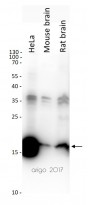
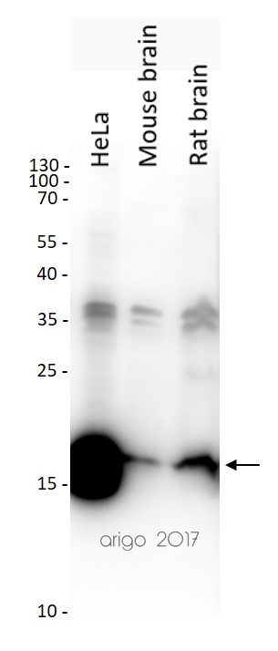
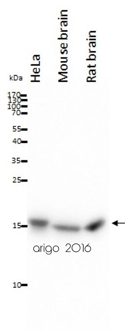
ARG65681 anti-Histone H3 antibody WB image
Western blot: 20 µg of HeLa, Mouse brain and Rat brain lysates stained with ARG65681 anti-Histone H3 antibody at 1:2000 dilution.
-
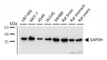
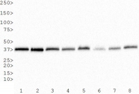
ARG10112 anti-GAPDH antibody [6C5] WB image
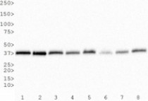
Western blot: 1) U87-MG 2) MCF-7 3) A549 4) DU145 5) SW480 6) rat brain 7) rat stomach 8) rat ovary stained with ARG10112 anti-GAPDH antibody [6C5] at 1:5000 dilution.
-
ARG10112 anti-GAPDH antibody [6C5] WB image
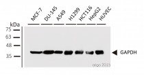
Western blot: 1) MCF-7 2) DU-145 3) A549 4) H1299 5) HCT116 6) HepG2 7) HUVEC stained with ARG10112 anti-GAPDH antibody [6C5] at 1:1000 dilution.
-

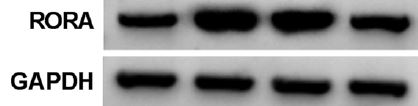
ARG10112 anti-GAPDH antibody [6C5] WB image
Western blot: PDLCs stained with ARG58393 anti-RORA antibody and ARG10112 anti-GAPDH antibody [6C5].
From Wang J et al. Arch Oral Biol- (2021), doi: 10.1016/j.archoralbio.2021.105053, Fig. 5. C.
-
ARG10112 anti-GAPDH antibody [6C5] WB image
Western Blot: 1) HeLa, 2) NTERA-2, 3) A-431, 4) HepG2, 5) MCF-7, 6) NIH 3T3, 7) PC-12 and 8) COS-7 whole cell lysates stained with anti-GAPDH antibody [6C5] (ARG10112)
-

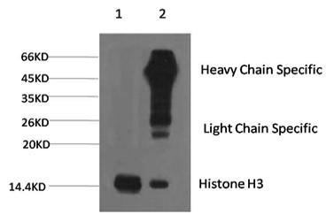
ARG65681 anti-Histone H3 antibody WB image
Western blot: 1) HeLa, 2) Raw, 3) Mouse brain tissue, and 4) Rat brain tissue lysates stained with ARG65681 anti-Histone H3 antibody at 1:5000 dilution.
-
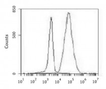
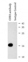
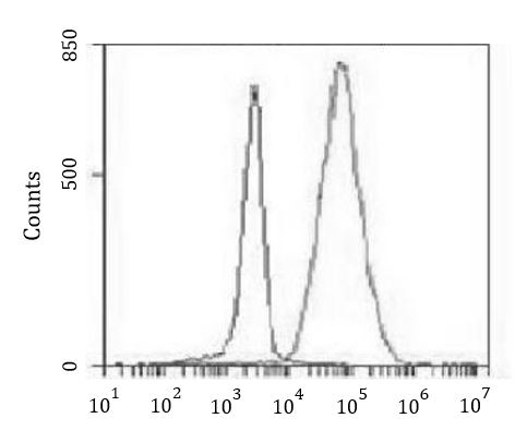
ARG54003 anti-COX4 antibody FACS image
Flow Cytometry: K562 cells stained with ARG54003 anti-COX4 antibody at 1:100 dilution (right histogram) or isotype control (left histogram), followed by incubation with FITC labelled secondary antibody.
-

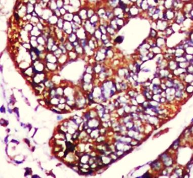
ARG54003 anti-COX4 antibody IHC-P image
Immunohistochemistry: Paraffin-embedded Human colorectal carcinoma stained with ARG54003 anti-COX4 antibody at 1:50 dilution. Antigen Retrieval: High-pressure and temperature Citrate buffer (pH 6.0).
-
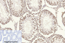
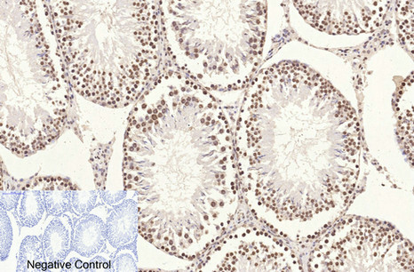
ARG65681 anti-Histone H3 antibody IHC-P image
Immunohistochemistry: Paraffin-embedded Rat testis tissue stained with ARG65681 anti-Histone H3 antibody at 1:200 dilution (4°C, overnight). Antigen Retrieval: Boil tissue section in Sodium citrate buffer (pH 6.0) for 20 min. Secondary antibody was diluted at 1:200 (RT, 30 min).
Negative control was used by secondary antibody only.
-
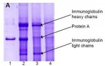
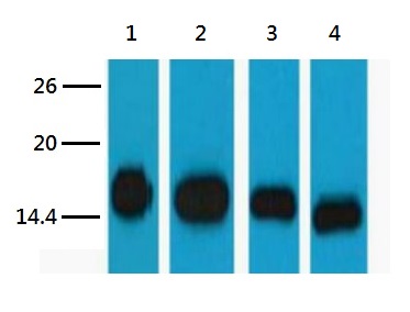
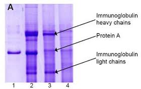
ARG10112 anti-GAPDH antibody [6C5] IP image
Immunoprecipitation and western blot: 1) GAPDH (1 μg). 2) GAPDH IP from rat heart tissue extract. 3) Only GAPDH preincubated with Protein A Sepharose. 4) Only Protein A Sepharose stained with ARG10112 GAPDH antibody [6C5].
Mixture of protein A-Sepharose with ARG10112 anti-GAPDH and tissue extract was incubated for 30 min at room temperature and precipitated by centrifugation. Pellet was washed with PBS, suspended in reducing electrophoresis sample buffer and heated for 5 minutes at 100ºC. After centrifugation supernatant was loaded on gel and proteins were separated by SDS electrophoresis. -

ARG65681 anti-Histone H3 antibody IP image
Immunoprecipitation: 1) HeLa cell lysate stained with ARG65681 anti-Histone H3 antibody and 2) IP product immunoprecipitated by ARG65681 anti-Histone H3 antibody at 1:200 dilution.
-
ARG54003 anti-COX4 antibody IP image
Immunoprecipitation: HeLa cell lysates were immunoprecipitated and stained with ARG54003 anti-COX4 antibody.
-

ARG54003 anti-COX4 antibody WB image
Western blot: 20 µg of HeLa, Mouse brain and Rat brain lysates stained with ARG54003 anti-COX4 antibody at 1:1000 dilution.
-
ARG54003 anti-COX4 antibody ICC/IF image
Immunofluorescence: 100% Methanol fixed (RT, 10 min) HeLa cells stained with ARG54003 anti-COX4 antibody (green) at 1:150 dilution.
Secondary antibody: ARG55393 Goat anti-Mouse IgG (H+L) antibody (FITC)
-

ARG10112 anti-GAPDH antibody [6C5] WB image
Western blot: pAFs, AcoSV40, and AcoSI-1 stained with ARG10112 anti-GAPDH antibody [6C5] at 1:5000 dilution.
From Michele N Dill et al. PLoS One. (2023), doi: 10.3389/fcell.2022.899869, Fig. 2. C.
-
ARG10112 anti-GAPDH antibody [6C5] WB image
Western blot: Porcine kidney stained with ARG10112 anti-GAPDH antibody [6C5].
From Jianni Huang et al. Front Cell Dev Biol (2022), doi: 10.3389/fcell.2022.899869, Fig. 2. E.
-

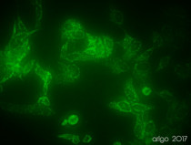
ARG10112 anti-GAPDH antibody [6C5] ICC/IF image
Immunofluorescence: 100% Methanol fixed (RT, 10 min) HeLa cells stained with ARG10112 anti-GAPDH antibody [6C5] (green) at 1:200 dilution.
Secondary antibody: ARG55393 Goat anti-Mouse IgG (H+L) antibody (FITC)
-
ARG10112 anti-GAPDH antibody [6C5] WB image
Western blot: Mouse samples stained with ARG10112 anti-GAPDH antibody [6C5] at 1:1000 dilution.
From Yun-Yun Li et al. Int J Biol Sci (2022), doi: 10.7150/ijbs.68224, Fig. 5. C.
-

ARG10112 anti-GAPDH antibody [6C5] WB image
Western blot: HUVEC stained with ARG10112 anti-GAPDH antibody [6C5].
From Bingzheng Lu et al. Oxid Med Cell Longev (2020), doi: 10.1155/2020/2048210, Fig. 5. B.
-
ARG65350 Goat anti-Mouse IgG antibody (HRP) IHC-P image
From Cheng-Feng Chu et al. J Pers Med. (2021), doi: 10.3390/jpm11121326, Fig. 6.
-
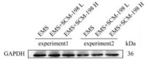
ARG65350 Goat anti-Mouse IgG antibody (HRP) WB image
Western blot: Neuro-2a stained with ARG22361 anti-Aquaporin 4 antibody , ARG40638 anti-MMP9 antibody and ARG65350 Goat anti-Mouse IgG antibody (HRP).
From Hung CY et al. Molecules (2022), doi: 10.3390/molecules27031066, Fig. 6A.
-
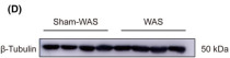
ARG65350 Goat anti-Mouse IgG antibody (HRP) WB image
Western blot: Rat basolateral amygdala stained with ARG62347 anti-beta Tubulin antibody [BT7R] at 1:1000 dilution, ARG65350 Goat anti-Mouse IgG antibody (HRP) at 1:5000 dilution.
From Guang-Bing Duan et al. CNS Neurosci Ther. (2024), doi: 10.1111/cns.14611, Fig. 4.D.


文献引用
KIF9 Ameliorates Neuropathology and Cognitive Dysfunction by Promoting Macroautophagy in a Mouse Model of Alzheimer's Disease
ARG10112: WB / Human, Mouse


A New Way to Engineer Cell Sheets for Articular Cartilage Regeneration
ARG65350; ICC/IF / Rabbit
Identification of RIPK3 as a target of flavonoids for anti-necroptosis in vitro
ARG10112; WB / Mouse
Disruption of BAG3-mediated BACE1 stabilization alleviates neuropathology and memory deficits in a mouse model of Alzheimer's disease
ARG10112; WB / Mouse
Structure-based design of potent and selective inhibitors targeting RIPK3 for eliminating on-target toxicity in vitro
ARG10112; WB / Human, Mouse
Alleviation of liver fibrosis by inhibiting a non-canonical ATF4-regulated enhancer program in hepatic stellate cells
ARG10112: WB / Human
Expression and correlation of Surfeit 4 gene in esophageal squamous cell carcinoma
ARG65350; WB /
PCSK6 exacerbates Alzheimer's disease pathogenesis by promoting MT5-MMP maturation
ARG10112: WB / Human, Mouse
Environmental acidification drives inter-organ energy mobilization to enhance reproductive performance in medaka (Oryzias latipes)
ARG65350; WB /
Biochanin A inhibits cardiac hypertrophy and fibrosis in vivo and in vitro
ARG10112: WB / Mouse, Rat
Shionone relieves oxygen-glucose deprivation/reoxygenation induced SH-SY5Y cells injury by inhibiting the p38 MAPK/NF-κB pathway
ARG10112: WB / Human
7,8-Dihydroxyflavone ameliorates cognitive impairment induced by repeated neonatal sevoflurane exposures in mice through increasing tau O-GlcNAcylation
ARG10112: WB / Mouse
The effect of long-term administration of green tea catechins on aging-related cardiac diastolic dysfunction and decline of troponin I
ARG10112: WB / Mouse
PPARγ activation suppresses chondrocyte ferroptosis through mitophagy in osteoarthritis
ARG10112: WB / Rat
Resveratrol inhibits Toxoplasma gondii-induced lung injury, inflammatory cascade and evidences of its mechanism of action
ARG10112: WB / Mouse
Knockdown of Porf-2 restores visual function after optic nerve crush injury
ARG10112: WB / Mouse
KDF1 Promoted Proliferation, Migration and Invasion of Lung Adenocarcinoma Cells through Activating STAT3 and AKT Pathway
ARG65350: WB /
Uric acid mitigates cognitive deficits via transcription factor EB - mediated microglial autophagy in mice models of Alzheimer's disease
ARG10112: WB / Mouse
黄芪甲苷预处理对大鼠肠缺血再灌注所致肺损伤的影响及其机制
ARG10112: WB / Rat
Discovery of the First Subnanomolar PPARα/δ Dual Agonist for the Treatment of Cholestatic Liver Diseases
ARG10112: WB / Mouse
Mesenchymal Stem Cells-Derived Exosomes Ameliorate Ischemia/Reperfusion Induced Acute Kidney Injury in a Porcine Model
ARG10112: WB / Pig
Lack of interferon regulatory factor 3 leads to anxiety/depression-like behaviors through disrupting the balance of neuronal excitation and inhibition in mice
ARG10112: WB / Mouse
Downregulation of lncRNA NEAT1 alleviates sepsis-induced acute kidney injury
ARG10112: WB / Human
Genetic labeling reveals cellular expression pattern of neuregulin 1 in mouse forebrain
ARG10112: WB / Human
Qi-Tai-Suan, an oleanolic acid derivative, ameliorates ischemic heart failure via suppression of cardiac apoptosis, inflammation and fibrosis
ARG10112: WB / Human
Gut microbiome dysbiosis contributes to abdominal aortic aneurysm by promoting neutrophil extracellular trap formation
ARG65681: IHC-P / Mouse
SCM-198 Prevents Endometriosis By Reversing Low Autophagy Levels Of Endometriotic Stromal Cells Via Inhibiting TNF-Α-Aromatase-Estrogen-Erα Pathway And Promoting PR Expression
ARG10122: WB / Human
SCM-198 Prevents Endometriosis by Reversing Low Autophagy of Endometrial Stromal Cell via Balancing ERα and PR Signals
ARG10112: WB / Mouse
Pharmacological inhibition of SMYD2 protects against cisplatin-induced acute kidney injury in mice
ARG10112: WB / Mouse
Evaluation of Class IIa Histone Deacetylases Expression and In Vivo Epigenetic Imaging in a Transgenic Mouse Model of Alzheimer's Disease
ARG10112: WB / Human
7,8-dihydroxyflavone ameliorates motor deficits via regulating autophagy in MPTP-induced mouse model of Parkinson's disease
ARG10112: WB / Mouse
M351-0056 is a novel low MW compound modulating the actions of the immune-checkpoint protein VISTA.
ARG10112: WB / Human, Mouse
MicroRNA-145 Delay Plaque Erosion by Down-Regulating TGFβr Ii and Suppressing Autophagy of Vascular Smooth Muscle Cells.
ARG65350: WB /
Cardiac-Specific Gene TNNI3 as a Potential Oncogene for Kidney Cancer and Its Involvement in Wnt Signaling Pathway.
ARG10112: WB / Human
Circ_0081572 inhibits the progression of periodontitis through regulating the miR-378h/RORA axis.
ARG10112: WB / Human
Inactivation of the tumor suppressor p53 by long noncoding RNA RMRP.
ARG65350: WB / Mouse
Excessive fibroblast growth factor 23 promotes renal fibrosis in mice with type 2 cardiorenal syndrome.
ARG10112: WB / Mouse, Rat
Design, Synthesis, and Activity Evaluation of Novel Acyclic Nucleosides as Potential Anticancer Agents In Vitro and In Vivo.
ARG65350: WB / Mouse
Blocking the CCL5-CCR5 Axis Using Maraviroc Promotes M1 Polarization of Macrophages Cocultured with Irradiated Hepatoma Cells.
ARG65350: WB / Mouse
Synthesis and anti-inflammatory activity of saponin derivatives of δ-oleanolic acid
ARG10112: WB / Human
Intermittent pressure imitating rolling manipulation ameliorates injury in skeletal muscle cells through oxidative stress and lipid metabolism signalling pathways.
ARG65350: WB / Mouse
Repurposing the FDA-approved anticancer agent ponatinib as a fluconazole potentiator by suppression of multidrug efflux and Pma1 expression in a broad spectrum of yeast species.
ARG65350: WB / Mouse
Structural Basis of VSIG3: The Ligand for VISTA.
ARG10112: WB / Human
Mesenchymal Stem Cell-Derived Exosomes Ameliorate Alzheimer's Disease Pathology and Improve Cognitive Deficits.
ARG10112: WB / Human
Curcumin attenuates renal ischemia reperfusion injury via JNK pathway with the involvement of p300/CBP-mediated histone acetylation
ARG10112: WB / Rat
The Phosphorylation Status of Drp1-Ser637 by PKA in Mitochondrial Fission Modulates Mitophagy via PINK1/Parkin to Exert Multipolar Spindles Assembly during Mitosis.
ARG10112: WB / Human
Decidual NR2F2-Expressing CD4 + T Cells Promote TH2 Transcriptional Program During Early Pregnancy.
ARG65350: WB /
Study of isolation and characterization of extracellular vesicles in senescent tumor cells induced by chemotherapeutics.
ARG10112: WB / Human
Increased expression levels of inflammatory cytokines and adhesion molecules in lipopolysaccharide‑induced acute inflammatory apoM‑/‑ mice
ARG10112: WB / Mouse
Discovery of AdipoRon analogues as novel AMPK activators without inhibiting mitochondrial complex I.
ARG10112: WB / Human, Mouse
CLC-3 and SOX2 regulate the cell cycle in DU145 cells
ARG65350: WB /
High expression of FUSE binding protein 1 in breast cancer stimulates cell proliferation and diminishes drug sensitivity.
ARG65350: WB /
Cardiac troponin I R193H mutant interacts with HDAC1 to repress phosphodiesterase 4D expression in cardiomyocytes
ARG10112: WB / Mouse
Prokaryotic expression and immunogenicity analysis of Clostridium difficile binary toxin A.
ARG65350: WB / Mouse
BCL7C suppresses ovarian cancer growth by inactivating mutant p53.
ARG65350: WB /
A single-component light sensor system allows highly tunable and direct activation of gene expression in bacterial cells.
ARG65350: WB / Mouse
Ubiquitin ligase DTX3 empowers mutant p53 to promote ovarian cancer development
ARG65350: WB /
ERK activation precedes Purkinje cell loss in mice with Spinocerebellar ataxia type 17.
ARG10112: WB / Mouse
Codelivery of Anti-PD-1 Antibody and Paclitaxel with Matrix Metalloproteinase and pH Dual-Sensitive Micelles for Enhanced Tumor Chemoimmunotherapy.
ARG65350: WB, IHC-P /
miR-765 Impairs Pancreatic β-cell Function by Targeting PDX1 in type 2 Diabetes.
ARG65350: WB /
SMARCB1 Promotes Ubiquitination and Degradation of NR4A3 via Direct Interaction Driven by ROS in Vascular Endothelial Cell Injury
ARG10112, ARG65350: WB / Human, Mouse, Rat
Down-regulation of HTR1A-modulated ACC activation contributes to stress-induced visceral hyperalgesia in rats.
ARG65350: WB / Mouse
SCM-198 protects endometrial stromal cells from oxidative damage through Bax/Bcl-2 and ERK signaling pathways.
ARG10112: WB / Human
Effects of perfluorooctane on the retina as a short-term and small amounts remnant in rabbits.
ARG65350: IHC-P / Mouse
Epigenetic regulation of phosphodiesterase 4d in restrictive cardiomyopathy mice with cTnI mutations.
ARG10112: WB / Mouse
Icariin suppresses cell cycle transition and cell migration in ovarian cancer cells.
ARG65350: WB / Mouse
Tumor-associated neutrophils induce EMT by IL-17a to promote migration and invasion in gastric cancer cells.
ARG10112: WB / Human
Genetic labeling reveals temporal and spatial expression pattern of D2 dopamine receptor in rat forebrain.
ARG10112: WB / Rat
Ubiquitin ligase TRIM71 suppresses ovarian tumorigenesis by degrading mutant p53.
ARG65350: WB, IHC-P / Mouse
AdipoRon prevents l-thyroxine or isoproterenol-induced cardiac hypertrophy through regulating the AMPK-related pathway.
ARG10112: WB / Rat
The effect of melatonin on cardio fibrosis in juvenile rats with pressure overload and deregulation of HDACs.
ARG10112: WB / Rat
Epigallocatechin-3 gallate prevents pressure overload-induced heart failure by up-regulating SERCA2a via histone acetylation modification in mice.
ARG10112: WB / Mouse
MIR4532 gene variant rs60432575 influences the expression of KCNJ11 and the sulfonylureas-stimulated insulin secretion.
ARG65350: WB / Mouse
Knockdown of LGALS12 inhibits porcine adipocyte adipogenesis via PKA-Erk1/2 signaling pathway
ARG65350: WB / Mouse
抗菌肽Sublancin增强小鼠获得性免疫的研究
ARG65350: ELISA / Mouse
Treatment of diabetic mice with A Combination of Ketogenic Diet and Aerobic Exercise via modulations of PPARs gene programs.
ARG65350: WB / Mouse
Hydrogen sulfide protects against endoplasmic reticulum stress and mitochondrial injury in nucleus pulposus cells and ameliorates intervertebral disc degeneration.
ARG10112: WB / Human
Cardiac Protection of Valsartan on Juvenile Rats with Heart Failure by Inhibiting Activity of CaMKII via Attenuating Phosphorylation
ARG10112: WB / Rat
Checking transfer efficiency and equal loading via qualitative optical way in western blotting.
ARG10112: WB / Human
Study on the inhibition of hyperthermic CO₂ pneumoperitoneum on the proliferation and migration of colon cancer cells and its mechanism.
ARG65350: WB /
Acute hyperglycemia suppresses left ventricular diastolic function and inhibits autophagic flux in mice under prohypertrophic stimulation.
ARG10112: WB / Mouse
Reciprocal regulation between O-GlcNAcylation and tribbles pseudokinase 2 (TRIB2) maintains transformative phenotypes in liver cancer cells.
ARG10112: WB / Human


