ARG30276
Cytochrome-C fractionation Antibody Panel (Cytochrome-C, COX IV, beta Actin)
内含物
| 货号 | 内含物名称 | 宿主克隆性 | 反应 | 应用 | 包装 |
|---|---|---|---|---|---|
| ARG62461 | anti-Cytochrome C antibody [6H2.B4] | Mouse mAb | Hu, Ms, Rat | FACS, ICC/IF, IP, WB | 50 μl |
| ARG54003 | anti-COX4 antibody | Mouse mAb | Hu, Ms, Rat, Goat, Hm, Mk | FACS, ICC/IF, IHC-P, IP, WB | 50 μl |
| ARG53987 | anti-beta Actin antibody | Mouse mAb | Arabi, Chk, C. reinhardtii, Cucumber, Dm, Fsh, Goat, Hm, Hu, Mk, Ms, P. pastoris, Pig, Rb, Rat, Rice, S. cerevisiae, Yeast, Zfsh | WB | 50 μl |
| ARG65350 | Goat anti-Mouse IgG antibody (HRP) | Goat pAb | Ms | ELISA, IHC-P, WB | 50 μl |
概述
| 产品描述 | Apoptosis is a process of programmed cell death that occurs in multicellular organism. Intracellular or extracellular death signals kick-start apoptosis signaling by releasing Cytochrome-C from mitochondria through BAX. The released Cytochrome-C binds to APAF-1 and cleave caspase 9. Finally, caspase-3 and PARP will be cleaved and the cells officially go into apoptosis and die. The cellular localization of Cytochrome-C often informs us of the trigger of apoptosis pathway. Hence, Cytochrome-C antibody together with COX IV (mitochondria marker) and beta Actin (cytoplasm marker) antibodies form a great package for the study of Cytochrome-C localization through cell fractionation. Nur-E-Lamal et al. 2004. JBC 279:24911-24914 |
|---|---|
| 靶点名称 | Cytochrome-C fractionation |
| 別名 | Cytochrome-C fractionation antibody; beta Actin antibody; COX4 antibody; Cytochrome C antibody |
属性
| 存放说明 | For continuous use, store undiluted antibody at 2-8°C for up to a week. For long-term storage, aliquot and store at -20°C or below. Storage in frost free freezers is not recommended. Avoid repeated freeze/thaw cycles. Suggest spin the vial prior to opening. The antibody solution should be gently mixed before use. |
|---|---|
| 注意事项 | For laboratory research only, not for drug, diagnostic or other use. |
生物信息
| 全名 | Antibody Panel for Cytochrome-C fractionation (Cytochrome-C, COX IV, beta Actin) |
|---|---|
| 产品亮点 | Related Product: anti-Cytochrome C antibody; |
检测图片 (13) Click the Picture to Zoom In
-
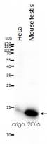
ARG62461 anti-Cytochrome C antibody [6H2.B4] WB image
Western blot: 20 µg of HeLa and Mouse testis lysates stained with ARG62461 anti-Cytochrome C antibody [6H2.B4] at 1:500 dilution.
-
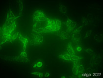
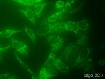
ARG54003 anti-COX4 antibody ICC/IF image
Immunofluorescence: 100% Methanol fixed (RT, 10 min) HeLa cells stained with ARG54003 anti-COX4 antibody (green) at 1:150 dilution.
Secondary antibody: ARG55393 Goat anti-Mouse IgG (H+L) antibody (FITC)
-
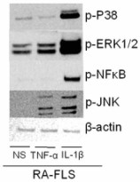
ARG53987 anti-beta Actin antibody WB image
Western blot: RA-FLSs stained with ARG53987 anti-beta Actin antibody.
From Machado CRL et al. Preprint- (2023), doi: 10.1186/s42358-022-00281-0, Fig. 5. E.
-
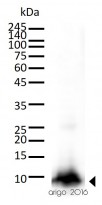
ARG62461 anti-Cytochrome C antibody [6H2.B4] WB image
Western blot: 30 µg of Mouse kidney lysate stained with ARG62461 anti-Cytochrome C antibody [6H2.B4] at 1:500 dilution.
-
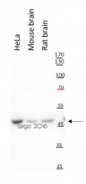
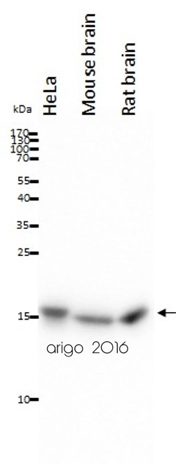
ARG53987 anti-beta Actin antibody WB image
Western blot: 20 µg of HeLa, Mouse brain and Rat brain lysates stained with ARG53987 anti-beta Actin antibody at 1:10000 dilution.
-
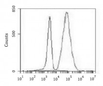
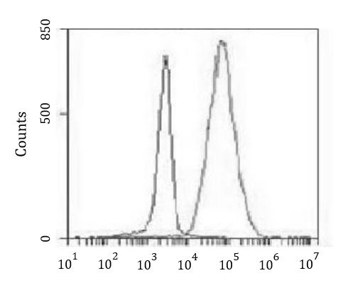
ARG54003 anti-COX4 antibody FACS image
Flow Cytometry: K562 cells stained with ARG54003 anti-COX4 antibody at 1:100 dilution (right histogram) or isotype control (left histogram), followed by incubation with FITC labelled secondary antibody.
-

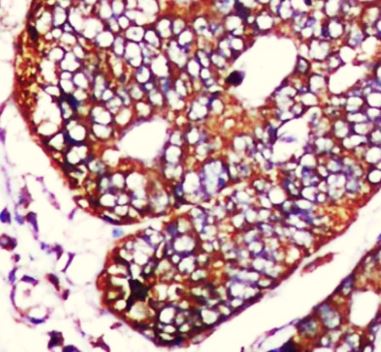
ARG54003 anti-COX4 antibody IHC-P image
Immunohistochemistry: Paraffin-embedded Human colorectal carcinoma stained with ARG54003 anti-COX4 antibody at 1:50 dilution. Antigen Retrieval: High-pressure and temperature Citrate buffer (pH 6.0).
-
ARG54003 anti-COX4 antibody IP image
Immunoprecipitation: HeLa cell lysates were immunoprecipitated and stained with ARG54003 anti-COX4 antibody.
-

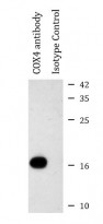
ARG54003 anti-COX4 antibody WB image
Western blot: 20 µg of HeLa, Mouse brain and Rat brain lysates stained with ARG54003 anti-COX4 antibody at 1:1000 dilution.
-
ARG54003 anti-COX4 antibody ICC/IF image
Immunofluorescence: 100% Methanol fixed (RT, 10 min) HeLa cells stained with ARG54003 anti-COX4 antibody (green) at 1:150 dilution.
Secondary antibody: ARG55393 Goat anti-Mouse IgG (H+L) antibody (FITC)
-
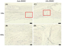
ARG65350 Goat anti-Mouse IgG antibody (HRP) IHC-P image
From Cheng-Feng Chu et al. J Pers Med. (2021), doi: 10.3390/jpm11121326, Fig. 6.
-
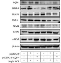
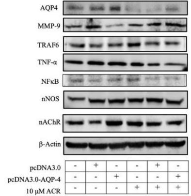
ARG65350 Goat anti-Mouse IgG antibody (HRP) WB image
Western blot: Neuro-2a stained with ARG22361 anti-Aquaporin 4 antibody , ARG40638 anti-MMP9 antibody and ARG65350 Goat anti-Mouse IgG antibody (HRP).
From Hung CY et al. Molecules (2022), doi: 10.3390/molecules27031066, Fig. 6A.
-
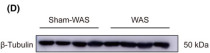
ARG65350 Goat anti-Mouse IgG antibody (HRP) WB image
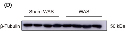
Western blot: Rat basolateral amygdala stained with ARG62347 anti-beta Tubulin antibody [BT7R] at 1:1000 dilution, ARG65350 Goat anti-Mouse IgG antibody (HRP) at 1:5000 dilution.
From Guang-Bing Duan et al. CNS Neurosci Ther. (2024), doi: 10.1111/cns.14611, Fig. 4.D.


文献引用
A New Way to Engineer Cell Sheets for Articular Cartilage Regeneration
ARG65350; ICC/IF / Rabbit


Expression and correlation of Surfeit 4 gene in esophageal squamous cell carcinoma
ARG65350; WB /
Environmental acidification drives inter-organ energy mobilization to enhance reproductive performance in medaka (Oryzias latipes)
ARG65350; WB /
KDF1 Promoted Proliferation, Migration and Invasion of Lung Adenocarcinoma Cells through Activating STAT3 and AKT Pathway
ARG65350: WB /
Morphofunctional analysis of fibroblast-like synoviocytes in human rheumatoid arthritis and mouse collagen-induced arthritis
ARG53987: WB / Human


