ARG66476
anti-RIPK1 / RIP1 phospho (Ser166) antibody [YJY-1-5]
anti-RIPK1 / RIP1 phospho (Ser166) antibody [YJY-1-5] for ICC/IF,IHC-Formalin-fixed paraffin-embedded sections,IHC-Frozen sections,Immunoprecipitation,Western blot and Human,Mouse,Rat

概述
| 产品描述 | Rabbit Monoclonal antibody [YJY-1-5] recognizes RIPK1 / RIP1 phospho (Ser166) |
|---|---|
| 反应物种 | Hu, Ms, Rat |
| 应用 | ICC/IF, IHC-Fr, IHC-P, IP, WB |
| 宿主 | Rabbit |
| 克隆 | Monoclonal |
| 克隆号 | YJY-1-5 |
| 同位型 | IgG |
| 靶点名称 | RIPK1 / RIP1 |
| 抗原物种 | Mouse |
| 抗原 | Phosphospecific peptide around Ser166 of Mouse RIPK1 / RIP1. |
| 偶联标记 | Un-conjugated |
| 別名 | Receptor-interacting protein 1; RIP-1; Receptor-interacting serine/threonine-protein kinase 1; RIP; Cell death protein RIP; RIP1; EC 2.7.11.1; Serine/threonine-protein kinase RIP |
应用说明
| 应用建议 |
|
||||||||||||
|---|---|---|---|---|---|---|---|---|---|---|---|---|---|
| 应用说明 | IHC-P: Antigen Retrieval: Citrate buffer (pH 6.0) at high pressure and temperature. * The dilutions indicate recommended starting dilutions and the optimal dilutions or concentrations should be determined by the scientist. |
属性
| 形式 | Liquid |
|---|---|
| 纯化 | Purification with Protein A. |
| 缓冲液 | PBS, 0.01% Sodium azide, 40% Glycerol and BSA. |
| 抗菌剂 | 0.01% Sodium azide |
| 稳定剂 | 40% Glycerol and BSA |
| 存放说明 | For continuous use, store undiluted antibody at 2-8°C for up to a week. For long-term storage, aliquot and store at -20°C. Storage in frost free freezers is not recommended. Avoid repeated freeze/thaw cycles. Suggest spin the vial prior to opening. The antibody solution should be gently mixed before use. |
| 注意事项 | For laboratory research only, not for drug, diagnostic or other use. |
生物信息
| 数据库连接 |
Swiss-port # Q13546 Human Receptor-interacting serine/threonine-protein kinase 1 Swiss-port # Q60855 Mouse Receptor-interacting serine/threonine-protein kinase 1 |
|---|---|
| 基因名称 | RIPK1 |
| 全名 | receptor (TNFRSF)-interacting serine-threonine kinase 1 |
| 背景介绍 | RIPK1 / RIP1 is a member of the receptor-interacting protein (RIP) family of serine/threonine protein kinases. The encoded protein plays a role in inflammation and cell death in response to tissue damage, pathogen recognition, and as part of developmental regulation. RIPK1/RIPK3 kinase-mediated necrosis is referred to as necroptosis. Genetic disruption of this gene in mice results in death shortly after birth. [provided by RefSeq, Aug 2017] |
| 生物功能 | RIPK1 / RIP1: Serine-threonine kinase which is a key regulator of both cell death and cell survival (PubMed:25459879). Exhibits kinase activity-dependent functions that trigger cell death and kinase-independent scaffold functions regulating inflammatory signaling and cell survival (PubMed:11101870, PubMed:25459879). Initiates ripoptocide which describes cell death that is dependent on RIPK1, be it apoptosis or necroptosis (PubMed:31457011). Upon binding of TNF to TNFR1, RIPK1 is recruited to the TNF-R1 signaling complex (TNF-RSC also known as complex I) where it acts as a scaffold protein promoting cell survival, in part, by activating the canonical NF-kB pathway. Specific conditions can however activate RIPK1, and its kinase activity then regulates assembly of two death-inducing complexes, namely complex IIa (RIPK1-FADD-CASP8) and the complex IIb (RIPK1-RIPK3-MLKL) and these complexes respectively drive apoptosis or necroptosis, a regulated form of necrosis (PubMed:19524513, PubMed:19524512, PubMed:29440439, PubMed:30988283). During embryonic development suppresses apoptosis and necroptosis and prevents the interaction of TRADD with FADD thereby limiting aberrant activation of CASP8. Phosphorylates DAB2IP at 'Ser-728' in a TNF- alpha-dependent manner, and thereby activates the MAP3K5-JNK apoptotic cascade (PubMed:17389591). Required for ZBP1-induced NF-kappaB activation and activation of NF-kappaB by DNA damage and IR. [UniProt] |
| 细胞定位 | Cytoplasm. Cell membrane. [UniProt] |
| 产品亮点 | Related products: RIPK1 antibodies; RIPK1 Duos / Panels; Anti-Rabbit IgG secondary antibodies; Related news: RIP1 activation and pathogenesis of NASH Ripoptosome & Necrosome antibody panels are launched RIP1 pSer166 antibody good for human and mouse samples |
| 预测分子量 | 76 kDa |
| 翻译后修饰 | Proteolytically cleaved by caspase-8 during TNF-induced apoptosis. Cleavage abolishes NF-kappa-B activation and enhances pro-apoptotic signaling through the TRADD-FADD interaction. RIPK1 and RIPK3 undergo reciprocal auto- and trans-phosphorylation. Phosphorylation of Ser-161 by RIPK3 is necessary for the formation of the necroptosis-inducing complex. Ubiquitinated by 'Lys-11'-, 'Lys-48'-, 'Lys-63'- and linear-linked type ubiquitin. Polyubiquitination with 'Lys-63'-linked chains by TRAF2 induces association with the IKK complex. Deubiquitination of 'Lys-63'-linked chains and polyubiquitination with 'Lys-48'-linked chains by TNFAIP3 leads to RIPK1 proteasomal degradation and consequently down-regulates TNF-alpha-induced NFkappa-B signaling. 'Lys-48'-linked polyubiquitination by RFFL or RNF34 also promotes proteasomal degradation and negatively regulates TNF-alpha-induced NFkappa-B signaling. Linear polyubiquitinated; the head-to-tail polyubiquitination is mediated by the LUBAC complex. LPS-mediated activation of NF-kappa-B. Also ubiquitinated with 'Lys-11'-linked chains. Polyubiquitinated with 'Lys-48' and 'Lys-63'-linked chains by BIRC2/c-IAP1 and BIRC3/c-IAP2, leading to activation of NF-kappa-B. [UniProt] |
检测图片 (7) Click the Picture to Zoom In
-

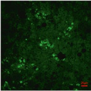
ARG66476 anti-RIPK1 / RIP1 phospho (Ser166) antibody [YJY-1-5] IHC-P image
Immunohistochemistry: Mouse sample stained with ARG66476 anti-RIPK1 / RIP1 phospho (Ser166) antibody [YJY-1-5].
From Hiroki Saijo et al. pre-print (2025), doi: 10.21203/rs.3.rs-5857284/v1, Fig. 6.
-
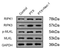
ARG66476 anti-RIPK1 / RIP1 phospho (Ser166) antibody [YJY-1-5] WB image
Western blot: HT22 stained with ARG41235 anti-RIPK3 / RIP3 phospho (Ser232) antibody and ARG66476 anti-RIPK1 / RIP1 phospho (Ser166) antibody [YJY-1-5].
From Lan-Lan Liu et al. Preprint (2024), doi: 10.21203/rs.3.rs-3841052/v1, Fig. 2A.
-

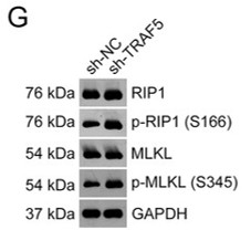
ARG66476 anti-RIPK1 / RIP1 phospho (Ser166) antibody [YJY-1-5] WB image
Western blot: HepG2 stained with ARG40183 anti-RIPK1 / RIP1 antibody and ARG66476 anti-RIPK1 / RIP1 phospho (Ser166) antibody [YJY-1-5].
From Wu G et al. PeerJ. (2023), doi: 10.7717/peerj.15551, Fig. 2. G.
-
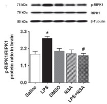
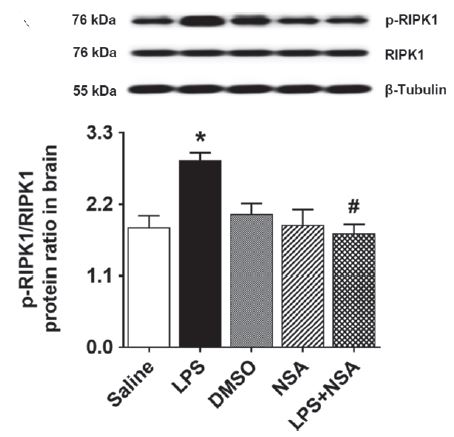
ARG66476 anti-RIPK1 / RIP1 phospho (Ser166) antibody [YJY-1-5] WB image
Western blot: Mouse brain stained with ARG55746 anti-RIPK1 / RIP1 antibody and ARG66476 anti-RIPK1 / RIP1 phospho (Ser166) antibody [YJY-1-5].
From Beyza Ozgen et al. Research Article (2022), doi: , Fig. 3. A.
-
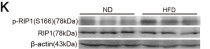
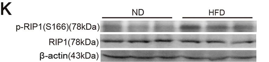
ARG66476 anti-RIPK1 / RIP1 phospho (Ser166) antibody [YJY-1-5] WB image
Western blot: Mouse liver stained with ARG66476 anti-RIPK1 / RIP1 phospho (Ser166) antibody [YJY-1-5].
From Tao L et al. Cell Death Differ- (2021), doi: 10.1038/s41418-020-00668-w, Fig. 1. K.
-

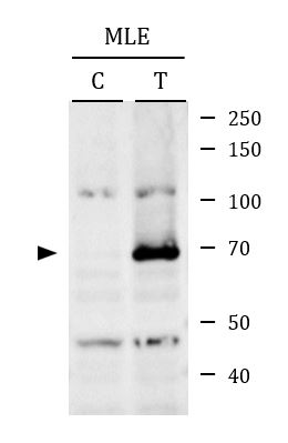
ARG66476 anti-RIPK1 / RIP1 phospho (Ser166) antibody [YJY-1-5] WB image
Western blot: MLE cells untreated control (C) or treated with 20 ng/ml of TNF alpha, 1 μM of BV-6 and 20 μM of Z-VAD-FMK for 8 hours (T). Cell lysates were stained with ARG66476 anti-RIPK1 / RIP1 phospho (Ser166) antibody [YJY-1-5].
-

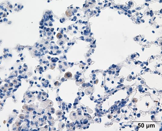
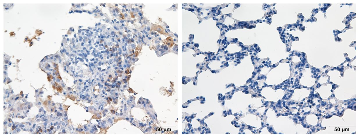
ARG66476 anti-RIPK1 / RIP1 phospho (Ser166) antibody [YJY-1-5] IHC-P image
Immunohistochemistry: Paraffin-embedded rat lung tissue from rat silicosis model. Antigen Retrieval: Citrate buffer (pH 6.0) at high pressure and temperature. The tissue section was stained with ARG66476 anti-RIPK1 / RIP1 phospho (Ser166) antibody [YJY-1-5] at 1:70 dilution. The picture on the right is negative control.


客户反馈
Excellent
anti-RIPK1 / RIP1 phospho (Ser166) antibody [YJY-1-5]
Application:WB
Sample:MLE cells untreated control (C) or treated with 20 ng/ml of TNF alpha, 1 μM of BV-6 and 20 μM of Z-VAD-FMK for 8 hours (T)
Primary Antibody Incubation Time:overnight
Primary Antibody Incubation Temperature:4 ºC

Excellent
anti-RIPK1 / RIP1 phospho (Ser166) antibody [YJY-1-5]
Application:IHC
Sample:Rat lung from rat silicosis model
Sample Preparation Method:Paraffin
Antigen Retrieval Buffer:Citrate acid buffer
Primary Antibody Dilution Factor:1:75
Excellent
anti-RIPK1 / RIP1 phospho (Ser166) antibody [YJY-1-5]
Application:IHC
Sample:Rat lung from rat silicosis model
Sample Preparation Method:Paraffin
Antigen Retrieval Buffer:Citrate buffer (pH 6.0)
Primary Antibody Dilution Factor:1:70
文献引用
Verifying inflammation and pancreatic blood flow in a mouse model of post-endoscopic retrograde cholangiopancreatography
IHC-P / Mouse


Inhibition of necroptosis mitigates paclitaxelinduced neuronal damage and cognitive impairment
WB / Mouse
Housing conditions affect enterocyte death mode and turnover rate in mouse small intestine
WB / Mouse
Silencing of TRAF5 enhances necroptosis in hepatocellular carcinoma by inhibiting LTBR-mediated NF-κB signaling
WB / Human
Neuronal MD2 induces long-term mental impairments in septic mice by facilitating necroptosis and apoptosis
WB / Mouse
Pyroptosis and Necroptosis Inhibitor Necrosulfonamide Prevents Lipopolysaccharide-Induced Inflammatory Hyperalgesia in Mice
WB / Mouse
NEK1-mediated retromer trafficking promotes blood-brain barrier integrity by regulating glucose metabolism and RIPK1 activation.
WB / Mouse
RIP1 kinase activity promotes steatohepatitis through mediating cell death and inflammation in macrophages.
WB, IHC-P / Mouse


