ARG64304
anti-PINK1 antibody
anti-PINK1 antibody for Flow cytometry,Western blot and Rat
Cell Biology and Cellular Response antibody; Metabolism antibody; Neuroscience antibody; Signaling Transduction antibody
概述
| 产品描述 | Goat Polyclonal antibody recognizes PINK1 |
|---|---|
| 反应物种 | Rat |
| 预测物种 | Hu |
| 应用 | FACS, WB |
| 宿主 | Goat |
| 克隆 | Polyclonal |
| 同位型 | IgG |
| 靶点名称 | PINK1 |
| 抗原物种 | Human |
| 抗原 | C-QGKAHLESRSYQEAQ |
| 偶联标记 | Un-conjugated |
| 別名 | PARK6; BRPK; PTEN-induced putative kinase protein 1; Serine/threonine-protein kinase PINK1, mitochondrial; EC 2.7.11.1 |
应用说明
| 应用建议 |
|
||||||
|---|---|---|---|---|---|---|---|
| 应用说明 | WB: Recommend incubate at RT for 1h. * The dilutions indicate recommended starting dilutions and the optimal dilutions or concentrations should be determined by the scientist. |
属性
| 形式 | Liquid |
|---|---|
| 纯化 | Purified from goat serum by antigen affinity chromatography. |
| 缓冲液 | Tris saline (pH 7.3), 0.02% Sodium azide and 0.5% BSA. |
| 抗菌剂 | 0.02% Sodium azide |
| 稳定剂 | 0.5% BSA |
| 浓度 | 0.5 mg/ml |
| 存放说明 | For continuous use, store undiluted antibody at 2-8°C for up to a week. For long-term storage, aliquot and store at -20°C or below. Storage in frost free freezers is not recommended. Avoid repeated freeze/thaw cycles. Suggest spin the vial prior to opening. The antibody solution should be gently mixed before use. |
| 注意事项 | For laboratory research only, not for drug, diagnostic or other use. |
生物信息
| 背景介绍 | This gene encodes a serine/threonine protein kinase that localizes to mitochondria. It is thought to protect cells from stress-induced mitochondrial dysfunction. Mutations in this gene cause one form of autosomal recessive early-onset Parkinson disease. [provided by RefSeq, Jul 2008] |
|---|---|
| 产品亮点 | Related products: PINK1 antibodies; Anti-Goat IgG secondary antibodies; Related news: Astrocyte-to-neuron conversion for Parkinson's disease treatment |
| 研究领域 | Cell Biology and Cellular Response antibody; Metabolism antibody; Neuroscience antibody; Signaling Transduction antibody |
| 预测分子量 | 63 kDa |
| 翻译后修饰 | Autophosphorylation at Ser-228 and Ser-402 is essential for Parkin/PRKN recruitment to depolarized mitochondria. Two shorter forms of 55 kDa and 48 kDa seem to be produced by proteolytic cleavage and localize mainly in cytosol. |
检测图片 (2) Click the Picture to Zoom In
-
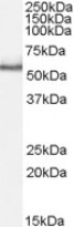
ARG64304 anti-PINK1 antibody WB image
Western Blot: Rat Testis lysate (35 µg protein in RIPA buffer) stained with ARG64304 anti-PINK1 antibody at 1 µg/ml dilution.
-

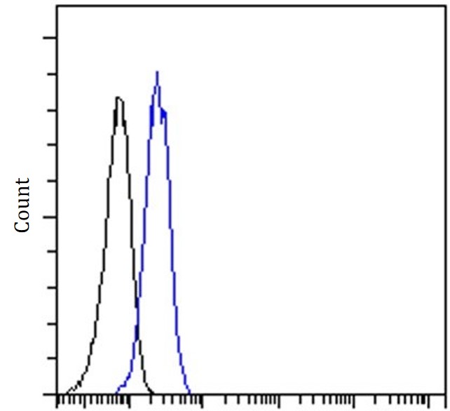
ARG64304 anti-PINK1 antibody FACS image
Flow Cytometry: Jurkat stained with ARG64304 anti-PINK1 antibody at 10 µg/ml dilution.