ARG59429
anti-MAVS antibody
anti-MAVS antibody for Flow cytometry,IHC-Formalin-fixed paraffin-embedded sections,Western blot and Human
概述
| 产品描述 | Rabbit Polyclonal antibody recognizes MAVS |
|---|---|
| 反应物种 | Hu |
| 应用 | FACS, IHC-P, WB |
| 宿主 | Rabbit |
| 克隆 | Polyclonal |
| 同位型 | IgG |
| 靶点名称 | MAVS |
| 抗原物种 | Human |
| 抗原 | Recombinant protein corresponding to L34-Q96 of Human MAVS. |
| 偶联标记 | Un-conjugated |
| 別名 | IPS1; IPS-1; Mitochondrial antiviral-signaling protein; Virus-induced-signaling adapter; VISA; CARDIF; Cardif; Interferon beta promoter stimulator protein 1; MAVS; Putative NF-kappa-B-activating protein 031N; CARD adapter inducing interferon beta |
应用说明
| 应用建议 |
|
||||||||
|---|---|---|---|---|---|---|---|---|---|
| 应用说明 | IHC-P: Antigen Retrieval: Heat mediated was performed in Citrate buffer (pH 6.0) for 20 min. * The dilutions indicate recommended starting dilutions and the optimal dilutions or concentrations should be determined by the scientist. |
属性
| 形式 | Liquid |
|---|---|
| 纯化 | Affinity purification with immunogen. |
| 缓冲液 | 0.9% NaCl, 0.2% Na2HPO4, 0.05% Sodium azide and 5% BSA. |
| 抗菌剂 | 0.05% Sodium azide |
| 稳定剂 | 5% BSA |
| 浓度 | 0.5 mg/ml |
| 存放说明 | For continuous use, store undiluted antibody at 2-8°C for up to a week. For long-term storage, aliquot and store at -20°C or below. Storage in frost free freezers is not recommended. Avoid repeated freeze/thaw cycles. Suggest spin the vial prior to opening. The antibody solution should be gently mixed before use. |
| 注意事项 | For laboratory research only, not for drug, diagnostic or other use. |
生物信息
| 数据库连接 |
Swiss-port # Q7Z434 Human Mitochondrial antiviral-signaling protein |
|---|---|
| 基因名称 | MAVS |
| 全名 | mitochondrial antiviral signaling protein |
| 背景介绍 | This gene encodes an intermediary protein necessary in the virus-triggered beta interferon signaling pathways. It is required for activation of transcription factors which regulate expression of beta interferon and contributes to antiviral immunity. Multiple transcript variants encoding different isoforms have been found for this gene. [provided by RefSeq, Sep 2011] |
| 生物功能 | Required for innate immune defense against viruses. Acts downstream of DDX58/RIG-I and IFIH1/MDA5, which detect intracellular dsRNA produced during viral replication, to coordinate pathways leading to the activation of NF-kappa-B, IRF3 and IRF7, and to the subsequent induction of antiviral cytokines such as IFN-beta and RANTES (CCL5). Peroxisomal and mitochondrial MAVS act sequentially to create an antiviral cellular state. Upon viral infection, peroxisomal MAVS induces the rapid interferon-independent expression of defense factors that provide short-term protection, whereas mitochondrial MAVS activates an interferon-dependent signaling pathway with delayed kinetics, which amplifies and stabilizes the antiviral response. May activate the same pathways following detection of extracellular dsRNA by TLR3. May protect cells from apoptosis. [UniProt] |
| 细胞定位 | Mitochondrion outer membrane. Mitochondrion. Peroxisome. [UniProt] |
| 产品亮点 | Related products: MAVS antibodies; Anti-Rabbit IgG secondary antibodies; Related news: Exploring Antiviral Immune Response |
| 预测分子量 | 57 kDa |
| 翻译后修饰 | Ubiquitinated (PubMed:19881509, PubMed:23087404). Undergoes 'Lys-48'-linked polyubiquitination catalyzed by ITCH; ITCH-dependent polyubiquitination is mediated by the interaction with PCBP2 and leads to MAVS/IPS1 proteasomal degradation (PubMed:19881509). Ubiquitinated by RNF125, leading to its degradation by the proteasome (PubMed:17460044). Undergoes 'Lys-48'-linked ubiquitination catalyzed by SMURF1 (PubMed:23087404). [UniProt] |
检测图片 (6) Click the Picture to Zoom In
-
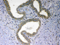
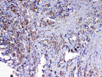
ARG59429 anti-MAVS antibody IHC-P image
Immunohistochemistry: Paraffin-embedded Human mammary cancer tissue. Antigen Retrieval: Heat mediated was performed in Citrate buffer (pH 6.0, epitope retrieval solution) for 20 min. The tissue section was blocked with 10% goat serum. The tissue section was then stained with ARG59429 anti-MAVS antibody at 1 µg/ml, overnight at 4°C.
-

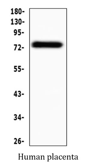
ARG59429 anti-MAVS antibody WB image
Western blot: 50 µg of sample under reducing conditions. Human placenta lysate stained with ARG59429 anti-MAVS antibody at 0.5 µg/ml dilution, overnight at 4°C.
-
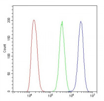
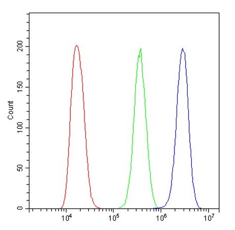
ARG59429 anti-MAVS antibody FACS image
Flow Cytometry: A431 cells were blocked with 10% normal goat serum and then stained with ARG59429 anti-MAVS antibody (blue) at 1 µg/10^6 cells for 30 min at 20°C, followed by incubation with DyLight®488 labelled secondary antibody. Isotype control antibody (green) was Rabbit IgG (1 µg/10^6) used under the same conditions. Unlabelled sample (red) was also used as a control.
-
ARG59429 anti-MAVS antibody IHC-P image
Immunohistochemistry: Paraffin-embedded Human lung cancer tissue. Antigen Retrieval: Heat mediated was performed in Citrate buffer (pH 6.0, epitope retrieval solution) for 20 min. The tissue section was blocked with 10% goat serum. The tissue section was then stained with ARG59429 anti-MAVS antibody at 1 µg/ml, overnight at 4°C.
-

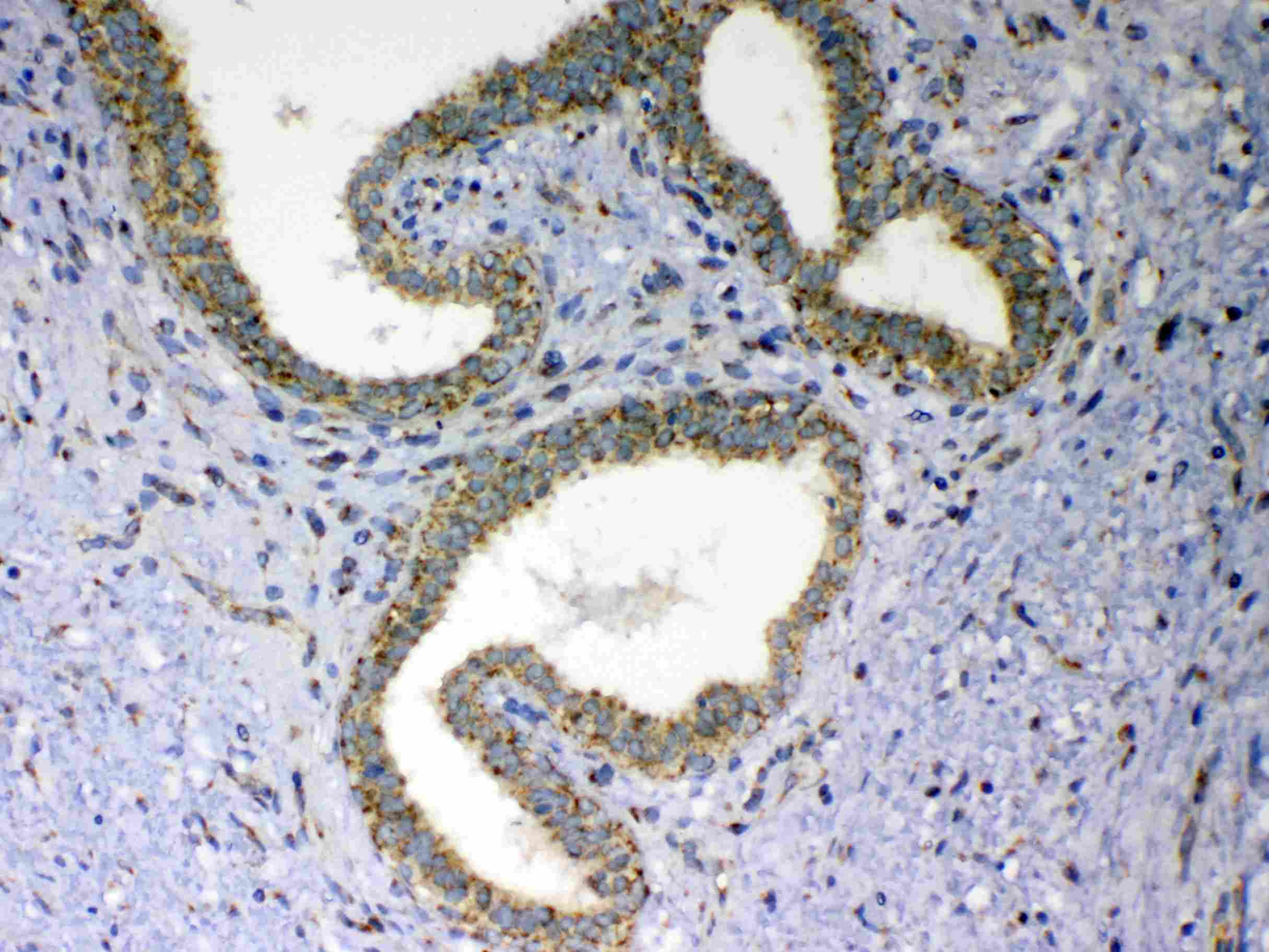
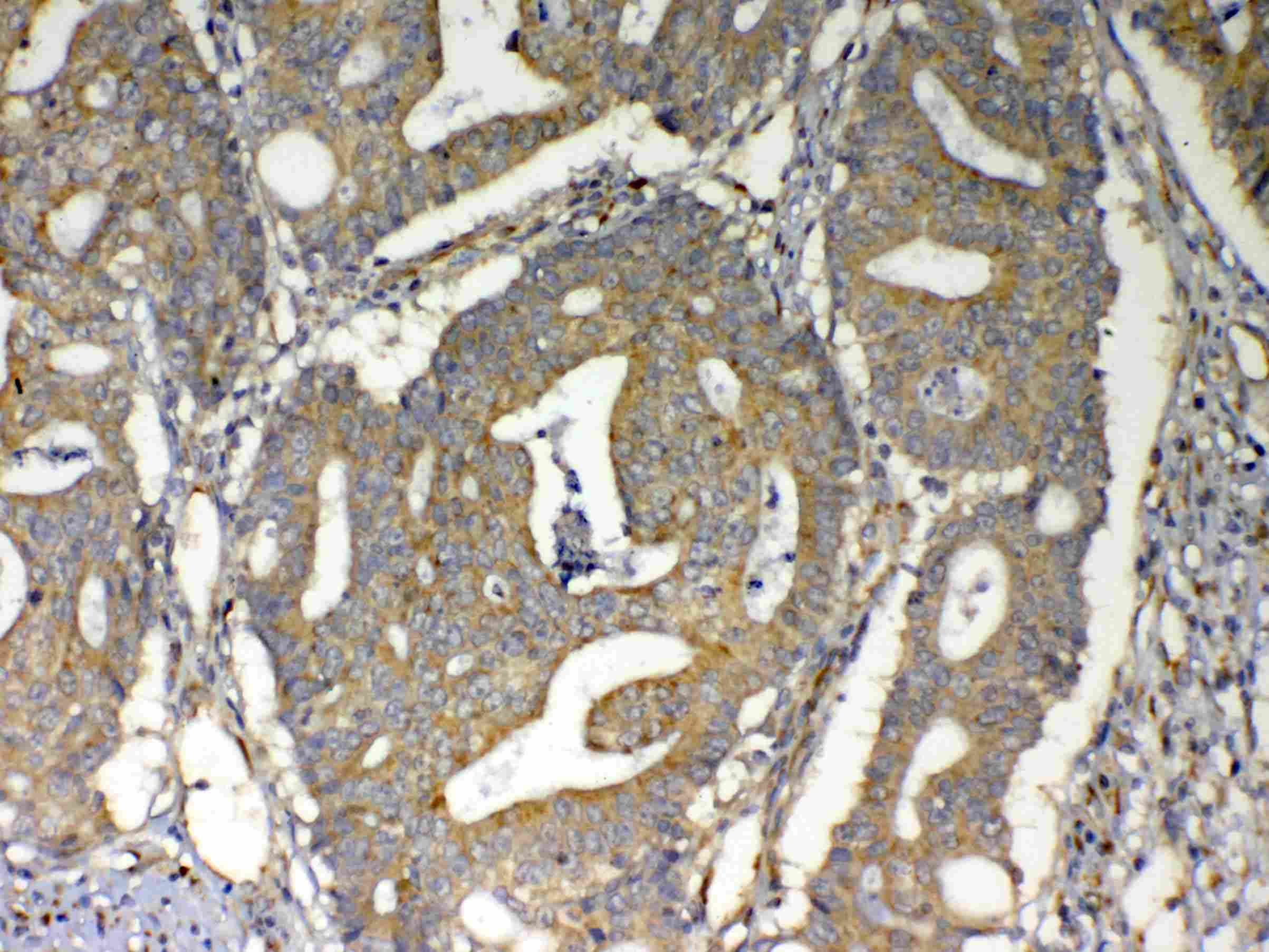
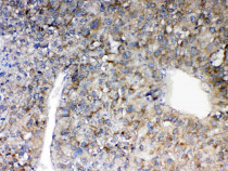
ARG59429 anti-MAVS antibody IHC-P image
Immunohistochemistry: Paraffin-embedded Human intestinal cancer tissue. Antigen Retrieval: Heat mediated was performed in Citrate buffer (pH 6.0, epitope retrieval solution) for 20 min. The tissue section was blocked with 10% goat serum. The tissue section was then stained with ARG59429 anti-MAVS antibody at 1 µg/ml, overnight at 4°C.
-
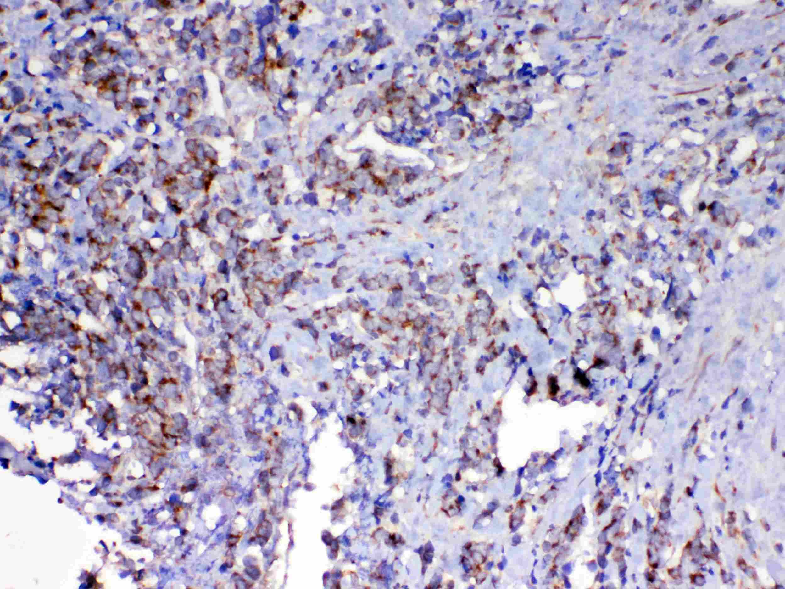
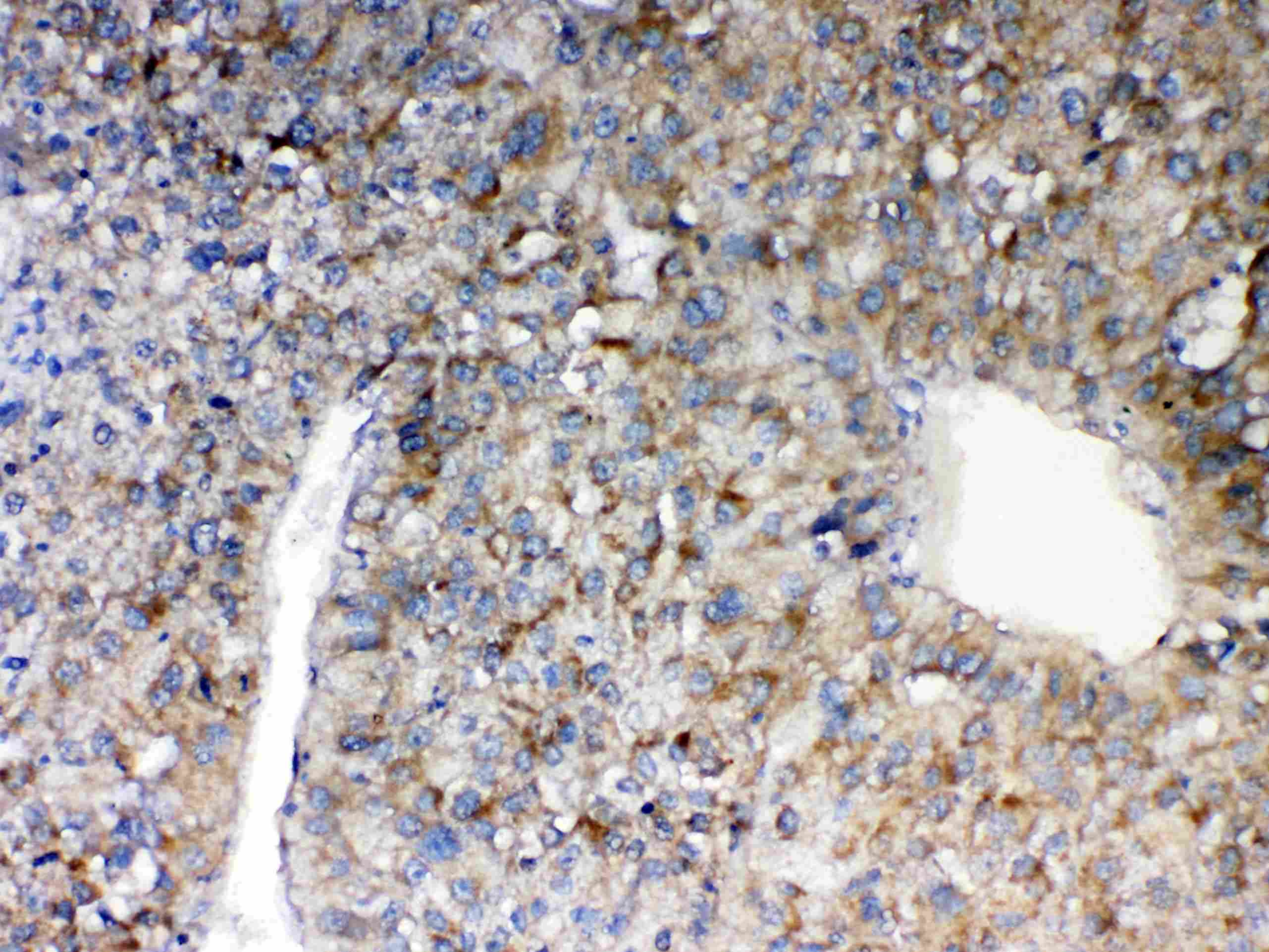
ARG59429 anti-MAVS antibody IHC-P image
Immunohistochemistry: Paraffin-embedded Human liver cancer tissue. Antigen Retrieval: Heat mediated was performed in Citrate buffer (pH 6.0, epitope retrieval solution) for 20 min. The tissue section was blocked with 10% goat serum. The tissue section was then stained with ARG59429 anti-MAVS antibody at 1 µg/ml, overnight at 4°C.