ARG56666
anti-IL1 alpha antibody
anti-IL1 alpha antibody for ELISA,IHC-Formalin-fixed paraffin-embedded sections,Neutralizing,Western blot and Human,Rat
概述
| 产品描述 | Rabbit Polyclonal antibody recognizes IL1 alpha |
|---|---|
| 反应物种 | Hu, Rat |
| 应用 | ELISA, IHC-P, Neut, WB |
| 宿主 | Rabbit |
| 克隆 | Polyclonal |
| 同位型 | IgG |
| 靶点名称 | IL1 alpha |
| 抗原物种 | Human |
| 抗原 | E.coli derived Recombinant Human IL-1α. (SAPFSFLSNV KYNFMRIIKY EFILNDALNQ SIIRANDQYL TAAALHNLDE AVKFDMGAYK SSKDDAKITV ILRISKTQLY VTAQDEDQPV LLKEMPEIPK TITGSETNLL FFWETHGTKN YFTSVAHPNL FIATKQDYWV CLAGGPPSIT DFQILENQA) |
| 偶联标记 | Un-conjugated |
| 別名 | IL-1 alpha; Interleukin-1 alpha; IL1; IL1F1; Hematopoietin-1; IL1-ALPHA; IL-1A |
应用说明
| 应用建议 |
|
||||||||||
|---|---|---|---|---|---|---|---|---|---|---|---|
| 应用说明 | * The dilutions indicate recommended starting dilutions and the optimal dilutions or concentrations should be determined by the scientist. |
属性
| 形式 | Liquid |
|---|---|
| 纯化 | Affinity purification with immunogen. |
| 缓冲液 | PBS (pH 7.2) |
| 浓度 | 1 mg/ml |
| 存放说明 | For continuous use, store undiluted antibody at 2-8°C for up to a week. For long-term storage, aliquot and store at -20°C or below. Storage in frost free freezers is not recommended. Avoid repeated freeze/thaw cycles. Suggest spin the vial prior to opening. The antibody solution should be gently mixed before use. |
| 注意事项 | For laboratory research only, not for drug, diagnostic or other use. |
生物信息
| 数据库连接 | |
|---|---|
| 基因名称 | IL1A |
| 全名 | interleukin 1, alpha |
| 背景介绍 | The protein encoded by this gene is a member of the interleukin 1 cytokine family. This cytokine is a pleiotropic cytokine involved in various immune responses, inflammatory processes, and hematopoiesis. This cytokine is produced by monocytes and macrophages as a proprotein, which is proteolytically processed and released in response to cell injury, and thus induces apoptosis. This gene and eight other interleukin 1 family genes form a cytokine gene cluster on chromosome 2. It has been suggested that the polymorphism of these genes is associated with rheumatoid arthritis and Alzheimer's disease. [provided by RefSeq, Jul 2008] |
| 生物功能 | Produced by activated macrophages, IL-1 stimulates thymocyte proliferation by inducing IL-2 release, B-cell maturation and proliferation, and fibroblast growth factor activity. IL-1 proteins are involved in the inflammatory response, being identified as endogenous pyrogens, and are reported to stimulate the release of prostaglandin and collagenase from synovial cells. [UniProt] |
| 产品亮点 | Related products: IL1 alpha antibodies; IL1 alpha ELISA Kits; IL1 alpha recombinant proteins; Anti-Rabbit IgG secondary antibodies; Related news: HMGB1 in inflammation Inflammatory Cytokines |
| 预测分子量 | 31 kDa |
检测图片 (6) Click the Picture to Zoom In
-
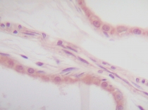
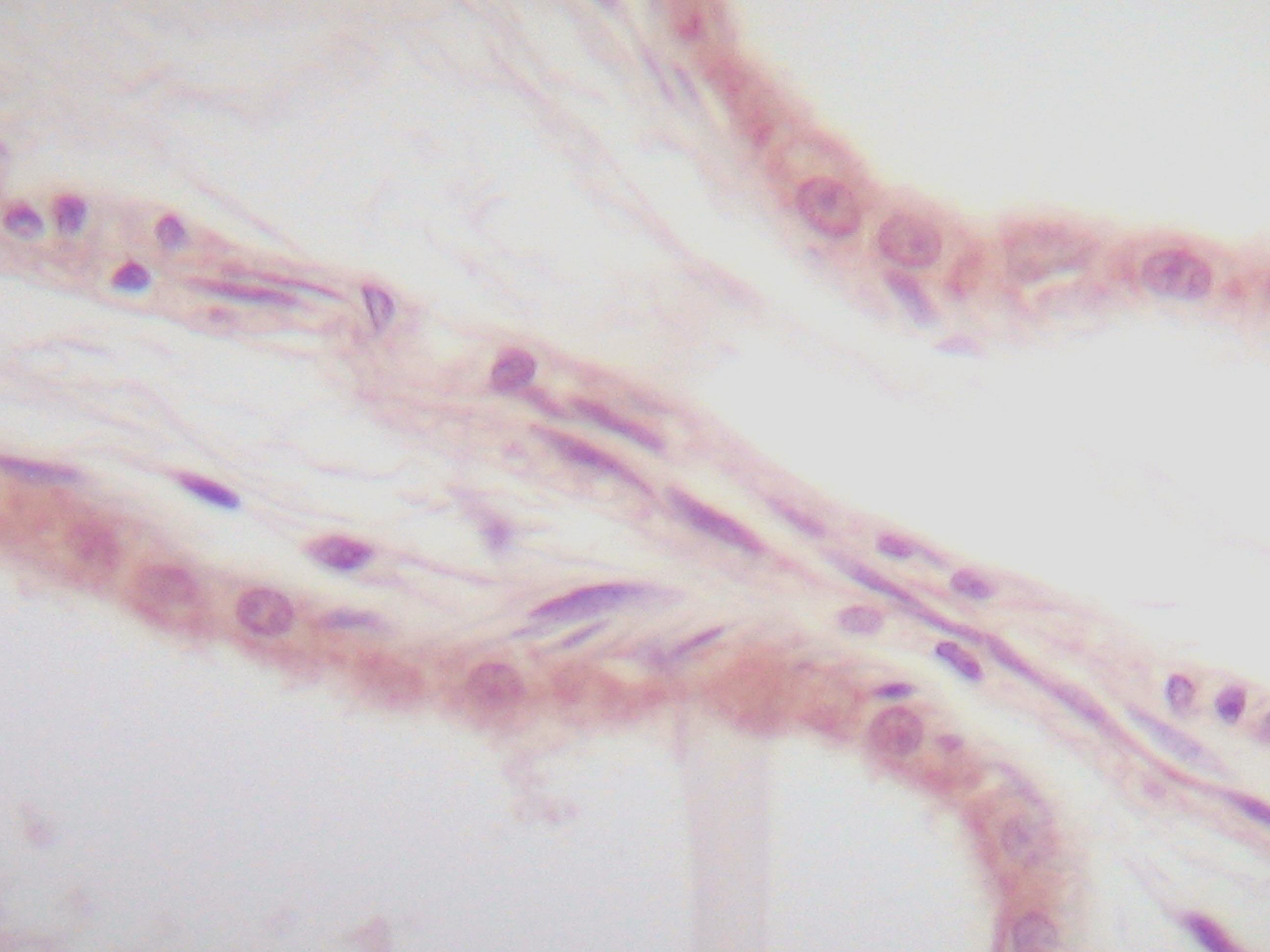
ARG56666 anti-IL1 alpha antibody IHC-P image
Immunohistochemistry: Formalin-fixed and paraffin-embedded sections of normal Human skin. The recommended ARG56666 anti-IL1 alpha antibody concentration is 1.0 µg/ml overnight at 4°C. An HRP-labeled polymer detection system was used with a non-alcohol soluble AEC chromogen. Antigen Retrieval: Incubate tissue section in a buffer (proteinase K) at RT for 10 min.
-
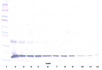
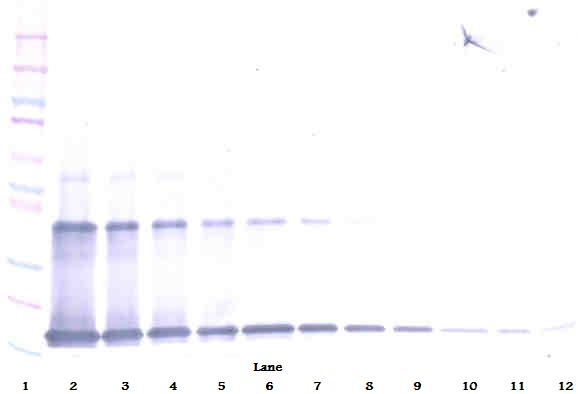
ARG56666 anti-IL1 alpha antibody WB image
Western blot: 250 - 0.24 ng of Human IL-1α stained with ARG56666 anti-IL1 alpha antibody, under reducing conditions.
-
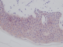
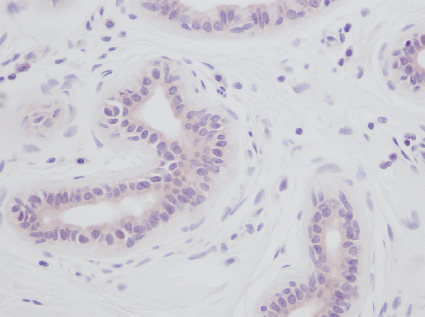
ARG56666 anti-IL1 alpha antibody IHC-P image
Immunohistochemistry: Formalin-fixed and paraffin-embedded sections of normal Human skin. The recommended ARG56666 anti-IL1 alpha antibody concentration is 1.0 µg/ml overnight at 4°C. An HRP-labeled polymer detection system was used with a non-alcohol soluble AEC chromogen. Antigen Retrieval: Incubate tissue section in a buffer (proteinase K) at RT for 10 min.
-
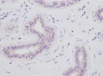
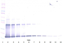
ARG56666 anti-IL1 alpha antibody WB image
Western blot: 250 - 0.24 ng of Human IL-1α stained with ARG56666 anti-IL1 alpha antibody, under non-reducing conditions.
-
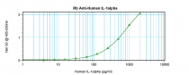
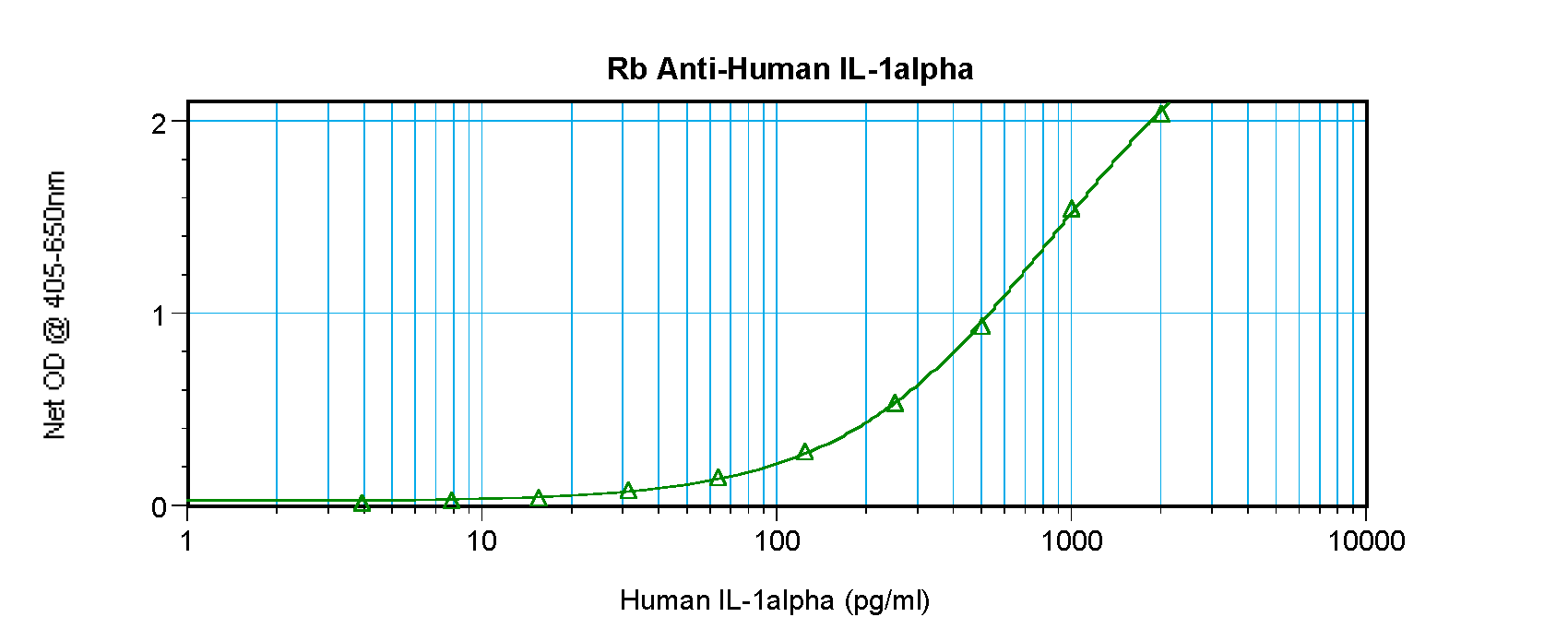
ARG56666 anti-IL1 alpha antibody standard curve image
Sandwich ELISA: ARG56666 anti-IL1 alpha antibody as a capture antibody at 0.5 - 2.0 µg/ml combined with ARG56776 anti-IL1 alpha antibody (Biotin) as a detection antibody. Results of a typical standard run with optical density.