ARG30324
Neuroinflammation Antibody Panel
内含物
| 货号 | 内含物名称 | 宿主克隆性 | 反应 | 应用 | 包装 |
|---|---|---|---|---|---|
| ARG11062 | anti-AIF1 / Iba 1 antibody | Rabbit pAb | Hu, Ms, Rat | ICC/IF, IHC-Fr, WB | 20 μl |
| ARG10514 | anti-CD68 antibody [KP1] | Mouse mAb | Hu, Rat | FACS, ICC/IF, IHC-P, WB | 20 μg |
| ARG52313 | anti-GFAP antibody | Chicken pAb | Bov, Cat, Chk, Hu, Ms, Rb, Rat, R. Mk | ELISA, FACS, ICC/IF, IHC-P, IHC-Fr, WB | 20 μl |
概述
| 产品描述 | Neuroinflammation Antibody Panel is an all-in-one solution to make neuroinflammation research easy and economic. It is ideal for studying neuroinflammation in rodent models and patient samples. This antibody panel comprises the most popular neuroinflammation markers: the active macroglial marker CD68 antibody, macroglial marker Iba-1 antibody, and astrocyte marker GFAP antibody. All the antibodies in this panel have excellent staining performance. Moreover, the different host species of the antibodies make the panel as the best choice for triple staining. Related news: Inflammation antibody panels are released Exploring Antiviral Immune Response |
|---|---|
| 靶点名称 | Neuroinflammation |
| 別名 | Neuroinflammation antibody; CD68 antibody; AIF1 / Iba 1 antibody; GFAP antibody |
属性
| 存放说明 | For continuous use, store undiluted antibody at 2-8°C for up to a week. For long-term storage, aliquot and store at -20°C or below. Storage in frost free freezers is not recommended. Avoid repeated freeze/thaw cycles. Suggest spin the vial prior to opening. The antibody solution should be gently mixed before use. |
|---|---|
| 注意事项 | For laboratory research only, not for drug, diagnostic or other use. |
生物信息
| 全名 | Antibody Panel for Neuroinflammation |
|---|---|
| 产品亮点 | Related products: anti-AIF1 / Iba 1 antibody; anti-CD68 antibody; anti-GFAP antibody; Inflammation antibodies; Inflammation Duos / Panels; |
检测图片 (11) Click the Picture to Zoom In
-

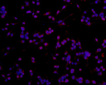
ARG10514 anti-CD68 antibody [KP1] ICC/IF image
Immunofluorescence: Human colon stained with ARG10514 anti-CD68 antibody [KP1].
From Li Y et al. Research Square (2022), doi: 10.3389/fmicb.2022.926915, Fig. 2C.
-
ARG11062 anti-AIF1 / Iba 1 antibody ICC/IF image (Customer's Feedback)
Immunofluorescence: BV-2 stained with ARG11062 anti-AIF1 / Iba 1 antibody at 1:100 dilution.
-
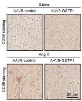
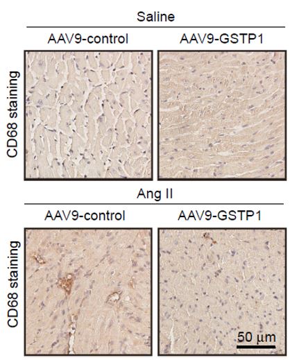
ARG10514 anti-CD68 antibody [KP1] IHC-P image
Immunohistochemistry: Mouse atrial stained with ARG10514 anti-CD68 antibody [KP1].
From Li H et al. Europace (2025), doi: 10.1093/europace/euaf083, Fig. 6E.
-

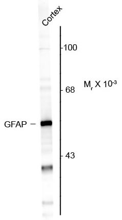
ARG52313 anti-GFAP antibody WB image
Western Blot: Rat cortex lysate showing specific immunolabeling of the ~50k GFAP protein stained with ARG52313 anti-GFAP antibody.
-
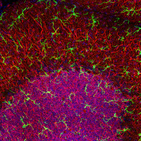
ARG11062 anti-AIF1 / Iba 1 antibody IHC-Fr image
Immunohistochemistry: High magnification stacked confocal image of Rat cerebellar molecular layer at top and granular layer below, stained with ARG11062 anti-AIF1 / Iba 1 antibody (green) at 1:1000 dilution. Nuclear DNA is shown with DAPI stain in blue.
Microglia are very small cells with fine processes spreading in three dimensions and so are best visualized in a confocal Z stack. Red shows the processes of Purkinje cells and the perikarya of granule cells revealed by an antibody to MAP2, at 1:5000 dilution.
-

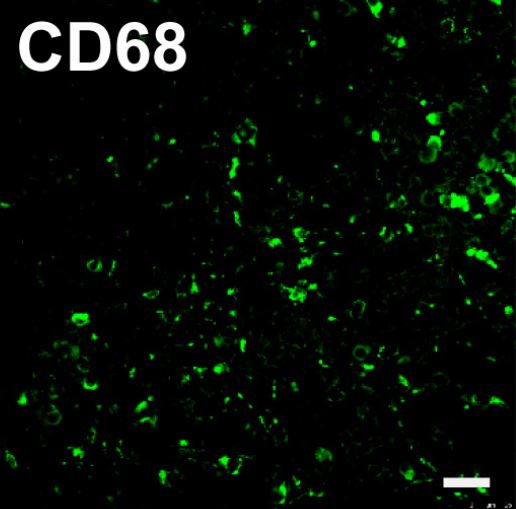
ARG10514 anti-CD68 antibody [KP1] IHC-P image
Immunohistochemistry: Formalin-fixed and paraffin-embedded Human tonsil stained with ARG10514 anti-CD68 antibody [KP1]. Antigen Retrieval: Boil tissue section in Citrate buffer (pH 6.0).
-

ARG52313 anti-GFAP antibody ICC/IF image
Immunofluorescence: Mixed cultures of neurons and glia stained with ARG52313 anti-GFAP antibody (red), and DNA (blue). Astrocytes stain strongly and specifically in a clearly filamentous fashion with this antibody.
-
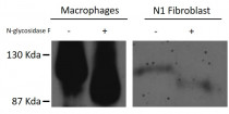
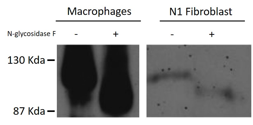
ARG10514 anti-CD68 antibody [KP1] WB image
Western blot: Human Macrophages and N1 Fibroblast untreated or treated with N-glycosidase F. The blots were stained with ARG10514 anti-CD68 antibody [KP1].
-
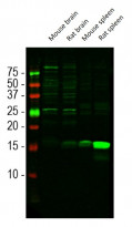
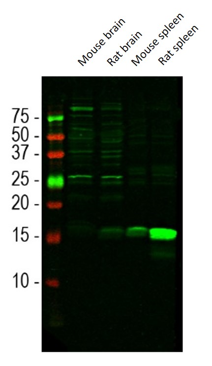
ARG11062 anti-AIF1 / Iba 1 antibody WB image
Western blot: Mouse brain, Rat brain, Mouse spleen, and Rat spleen lysates stained with ARG11062 anti-AIF1 / Iba 1 antibody at 1:1000 dilution.
Iba1 is a relatively minor protein of brain and is much more abundant in spleen, so the 15 kDa band is less obvious in CNS lysates. The other bands seen in the CNS lysates are of unknown origin but do not appear to compromise the migroglial specific staining seen with this antibody.
-

ARG10514 anti-CD68 antibody [KP1] FACS image
Flow Cytometry: THP1 cells prefixed with 4% PFA and then permeabilised with 0.25% saponin. Cells were stained with ARG10514 anti-CD68 antibody [KP1] (white area) or isotype control antibody (gray area).
-

ARG10514 anti-CD68 antibody [KP1] IHC-P image
Immunohistochemistry: Formalin-fixed and paraffin-embedded Human breast carcinoma stained with ARG10514 anti-CD68 antibody [KP1].


文献引用
Exploring macrophage polarization as a prognostic indicator for colorectal cancer
ARG10514; IHC-P / Human


GSTP1 inhibits angiotensin II-induced atrial fibrillation by regulating ferroptosis
ARG10514; IHC-P / Mouse
Hierarchical Assembly and Dynamic Reinforced Woven Biotextiles for Anisotropic and Viscoelastic Tendon Tissue Regeneration
ARG10514; IHC-P / Mouse
Mac-1 deficiency ameliorates pressure overloaded heart failure through inhibiting macrophage polarization and activation
ARG10514: IHC-Fr / Mouse
Cardamonin intervenes in myocardial hypertrophy progression by regulating Usp18
ARG10514: IHC-P / Mouse
Identification and experimental verification of immune-related hub genes in intervertebral disc degeneration
ARG10514: ICC/IF / Human
Aquaporin-9 Aggravates Lipopolysaccharides Induced Acute Lung Injury Via Facilitating M1-Like Macrophage Polarization
ARG10514: IHC-P / Mouse
Facile and rapid fabrication of a novel 3D-printable, visible light-crosslinkable and bioactive polythiourethane for large-to-massive rotator cuff tendon repair
ARG10514: IHC-P / Mouse
The Proinflammatory Role of Guanylate-Binding Protein 5 in Inflammatory Bowel Diseases
ARG10514: ICC/IF / Mouse
Complement-mediated M2/M1 macrophage polarization may be involved in crescent formation in lupus nephritis
ARG10514: IHC-P / Human
Btk knockout attenuates the liver inflammation in STZ-induced diabetic mice by suppressing NLRP3 inflammasome activation.
ARG10514: IHC-P / Mouse
Enhanced Bruton's tyrosine kinase activity in the kidney of patients with IgA nephropathy.
ARG10514: IHC-P / Human
Dark Sweet Cherry ( Prunus avium) Phenolics Enriched in Anthocyanins Induced Apoptosis in MDA-MB-453 Breast Cancer Cells through MAPK-Dependent Signaling and Reduced Invasion via Akt and PLCγ-1 Downregulation.
ARG11062: WB / Human
TAB1 regulates glycolysis and activation of macrophages in diabetic nephropathy.
ARG10514: IHC-P / Mouse
Exosomes from high glucose-treated macrophages activate glomerular mesangial cells via TGF-β1/Smad3 pathway in vivo and in vitro.
ARG10514: IHC-P / Mouse


