ARG44767
anti-Villin antibody
anti-Villin antibody for IHC-Formalin-fixed paraffin-embedded sections,Immunoprecipitation,Western blot and Human
概述
| 产品描述 | Mouse Monoclonal antibody recognizes Villin |
|---|---|
| 反应物种 | Hu |
| 应用 | IHC-P, IP, WB |
| 宿主 | Mouse |
| 克隆 | Monoclonal |
| 同位型 | IgG1 |
| 靶点名称 | Villin |
| 抗原物种 | Human |
| 偶联标记 | Un-conjugated |
| 別名 | Villin-1; D2S1471; VIL |
应用说明
| 应用建议 |
|
||||||||
|---|---|---|---|---|---|---|---|---|---|
| 应用说明 | * The dilutions indicate recommended starting dilutions and the optimal dilutions or concentrations should be determined by the scientist. |
属性
| 形式 | Liquid |
|---|---|
| 纯化 | Protein A purification |
| 缓冲液 | PBS with 0.09% sodium azide |
| 存放说明 | For continuous use, store undiluted antibody at 2-8°C for up to a week. For long-term storage, aliquot and store at -20°C or below. Storage in frost free freezers is not recommended. Avoid repeated freeze/thaw cycles. Suggest spin the vial prior to opening. The antibody solution should be gently mixed before use. |
| 注意事项 | For laboratory research only, not for drug, diagnostic or other use. |
生物信息
| 数据库连接 | |
|---|---|
| 基因名称 | VIL1 |
| 全名 | villin 1 |
| 背景介绍 | This gene encodes a member of a family of calcium-regulated actin-binding proteins. This protein represents a dominant part of the brush border cytoskeleton which functions in the capping, severing, and bundling of actin filaments. Two mRNAs of 2.7 kb and 3.5 kb have been observed; they result from utilization of alternate poly-adenylation signals present in the terminal exon. [provided by RefSeq, Jul 2008] |
| 生物功能 | E3 ubiquitin-protein ligase that is an intermolecular hub protein in the cell cycle network. Through cooperative DNA and histone binding, may contribute to a tighter epigenetic control of gene expression in differentiated cells. Ubiquitinates cyclins, CCND1 and CCNE1, in an apparently phosphorylation-independent manner and induces G1 arrest. Also ubiquitinates PCNP leading to its degradation by the proteasome. E3 SUMO-, but not ubiquitin-, protein ligase for ZNF131. [UniProt] |
| 细胞定位 | Cytoplasm, cytoskeleton. Cell projection, lamellipodium. Cell projection, ruffle. Cell projection, microvillus. Cell projection, filopodium tip. Cell projection, filopodium. Note=Relocalized in the tip of cellular protrusions and filipodial extensions upon infection with S.flexneri in primary intestinal epithelial cells (IEC) and in the tail-like structures forming the actin comets of S.flexneri. Redistributed to the leading edge of hepatocyte growth factor (HGF)-induced lamellipodia. [UniProt] |
| 预测分子量 | 93 kDa |
| 翻译后修饰 | Tyrosine phosphorylation is induced by epidermal growth factor (EGF) and stimulates cell migration (By similarity). Phosphorylated on tyrosine residues by SRC. The unphosphorylated form increases the initial rate of actin-nucleating activity, whereas the tyrosine-phosphorylated form inhibits actin-nucleating activity, enhances actin-bundling activity and enhances actin-severing activity by reducing high Ca(2+) requirements. The tyrosine-phosphorylated form does not regulate actin-capping activity. Tyrosine phosphorylation is essential for cell migration: tyrosine phosphorylation sites in the N-terminus half regulate actin reorganization and cell morphology, whereas tyrosine phosphorylation sites in the C-terminus half regulate cell migration via interaction with PLCG1. [UniProt] |
检测图片 (3) Click the Picture to Zoom In
-

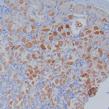
ARG44767 anti-Villin antibody IHC-P image
Immunohistochemistry: Human stomach stained with ARG44767 anti-Villin antibody at 4 µg/mL dilution.
-
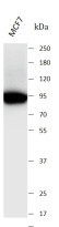
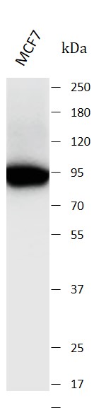
ARG44767 anti-Villin antibody WB image
Western blot: Caco2 stained with ARG44767 anti-Villin antibody at 1 µg/mL dilution.
-
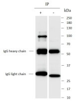
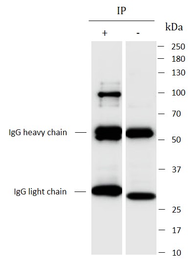
ARG44767 anti-Villin antibody IP image
Immunoprecipitation: Caco2 lysate immunoprecipitated with 2.5 µg of ARG44767 anti-Villin antibody.