ARG40764
anti-VE Cadherin antibody
anti-VE Cadherin antibody for Flow cytometry,ICC/IF,IHC-Frozen sections,IHC-Formalin-fixed paraffin-embedded sections,Western blot and Human
概述
| 产品描述 | Rabbit Polyclonal antibody recognizes VE Cadherin |
|---|---|
| 反应物种 | Hu |
| 预测物种 | Ms, Rat |
| 应用 | FACS, ICC/IF, IHC-Fr, IHC-P, WB |
| 宿主 | Rabbit |
| 克隆 | Polyclonal |
| 同位型 | IgG |
| 靶点名称 | VE Cadherin |
| 抗原物种 | Human |
| 抗原 | Recombinant protein corresponding to D48-R272 of Human VE Cadherin. |
| 偶联标记 | Un-conjugated |
| 別名 | 7B4 antigen; 7B4; Cadherin-5; VE-cadherin; CD144; CD antigen CD144; Vascular endothelial cadherin |
应用说明
| 应用建议 |
|
||||||||||||
|---|---|---|---|---|---|---|---|---|---|---|---|---|---|
| 应用说明 | IHC-P: Antigen Retrieval: Heat mediation was performed in Citrate buffer (pH 6.0, epitope retrieval solution) for 20 min. * The dilutions indicate recommended starting dilutions and the optimal dilutions or concentrations should be determined by the scientist. |
属性
| 形式 | Liquid |
|---|---|
| 缓冲液 | 0.2% Na2HPO4, 0.9% NaCl, 0.05% Sodium azide and 4% Trehalose. |
| 抗菌剂 | 0.05% Sodium azide |
| 稳定剂 | 4% Trehalose |
| 浓度 | 0.5 mg/ml |
| 存放说明 | For continuous use, store undiluted antibody at 2-8°C for up to a week. For long-term storage, aliquot and store at -20°C or below. Storage in frost free freezers is not recommended. Avoid repeated freeze/thaw cycles. Suggest spin the vial prior to opening. The antibody solution should be gently mixed before use. |
| 注意事项 | For laboratory research only, not for drug, diagnostic or other use. |
生物信息
| 数据库连接 | |
|---|---|
| 基因名称 | CDH5 |
| 全名 | cadherin 5, type 2 (vascular endothelium) |
| 背景介绍 | This gene is a classical cadherin from the cadherin superfamily and is located in a six-cadherin cluster in a region on the long arm of chromosome 16 that is involved in loss of heterozygosity events in breast and prostate cancer. The encoded protein is a calcium-dependent cell-cell adhesion glycoprotein comprised of five extracellular cadherin repeats, a transmembrane region and a highly conserved cytoplasmic tail. Functioning as a classic cadherin by imparting to cells the ability to adhere in a homophilic manner, the protein may play an important role in endothelial cell biology through control of the cohesion and organization of the intercellular junctions. An alternative splice variant has been described but its full length sequence has not been determined. [provided by RefSeq, Jul 2008] |
| 生物功能 | Cadherins are calcium-dependent cell adhesion proteins. They preferentially interact with themselves in a homophilic manner in connecting cells; cadherins may thus contribute to the sorting of heterogeneous cell types. This cadherin may play a important role in endothelial cell biology through control of the cohesion and organization of the intercellular junctions. It associates with alpha-catenin forming a link to the cytoskeleton. Acts in concert with KRIT1 to establish and maintain correct endothelial cell polarity and vascular lumen. These effects are mediated by recruitment and activation of the Par polarity complex and RAP1B. Required for activation of PRKCZ and for the localization of phosphorylated PRKCZ, PARD3, TIAM1 and RAP1B to the cell junction. [UniProt] |
| 细胞定位 | Cell junction. Cell membrane; Single-pass type I membrane protein. Note=Found at cell-cell boundaries and probably at cell-matrix boundaries. KRIT1 and CDH5 reciprocally regulate their localization to endothelial cell-cell junctions. [UniProt] |
| 预测分子量 | 88 kDa (unmodified); 90 - 140 kDa (glycosylated) |
| 翻译后修饰 | Phosphorylated on tyrosine residues by KDR/VEGFR-2. Dephosphorylated by PTPRB (By similarity). O-glycosylated. [UniProt] |
检测图片 (8) Click the Picture to Zoom In
-
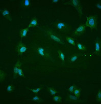
ARG40764 anti-VE Cadherin antibody ICC/IF image
Immunofluorescence: A549 cells were blocked with 10% goat serum and then stained with ARG40764 anti-VE Cadherin antibody (green) at 4 μg/ml dilution, overnight at 4°C. DAPI (blue) for nuclear staining.
-
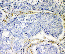
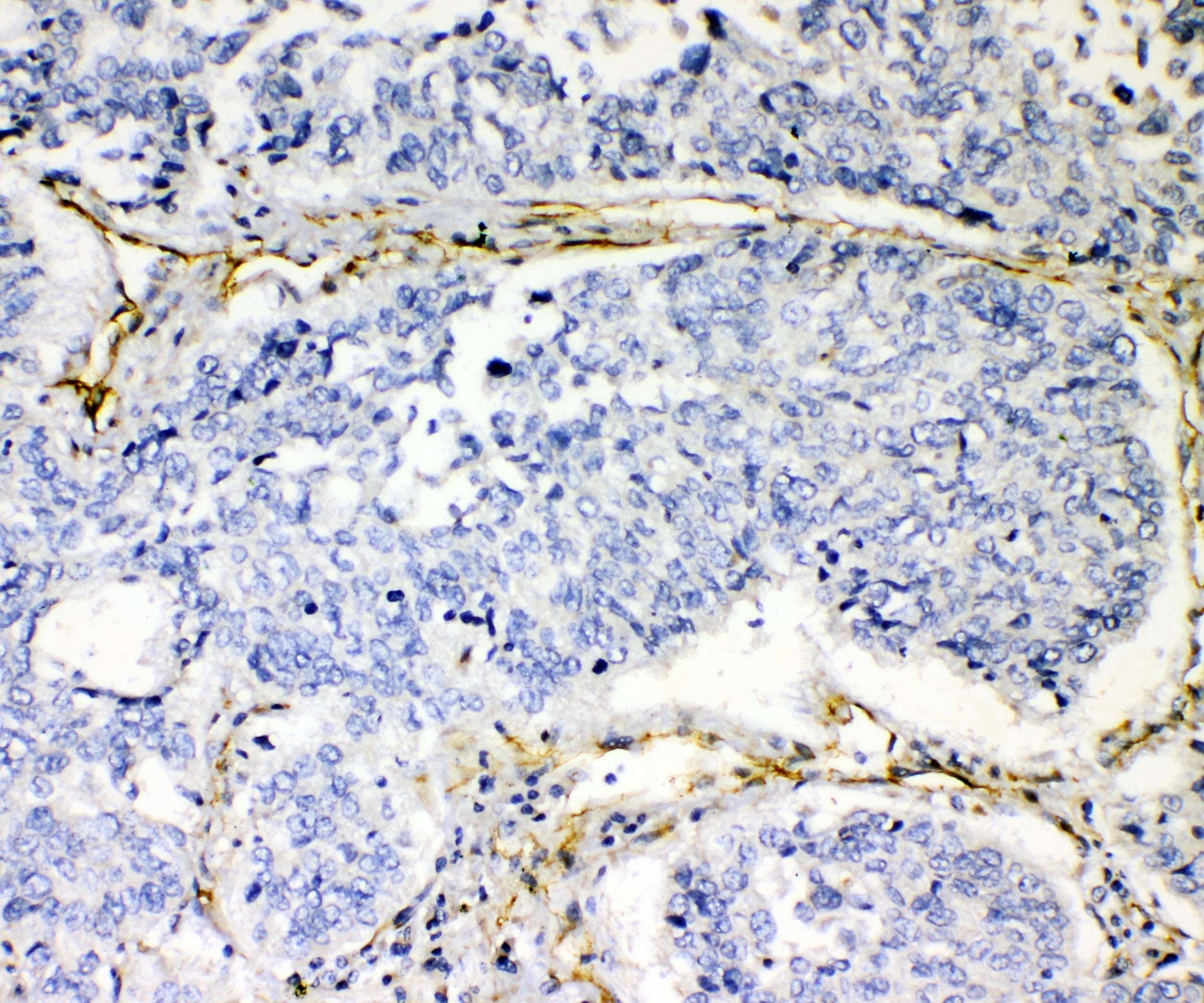
ARG40764 anti-VE Cadherin antibody IHC-P image
Immunohistochemistry: Paraffin-embedded Human lung cancer tissue. Antigen Retrieval: Heat mediation was performed in Citrate buffer (pH 6.0, epitope retrieval solution) for 20 min. The tissue section was blocked with 10% goat serum. The tissue section was then stained with ARG40764 anti-VE Cadherin antibody at 1 µg/ml, overnight at 4°C.
-
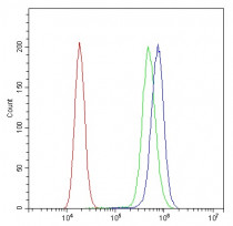
ARG40764 anti-VE Cadherin antibody FACS image
Flow Cytometry: A431 cells were blocked with 10% normal goat serum and then stained with ARG40764 anti-VE Cadherin antibody (blue) at 1 μg/10^6 cells for 30 min at 20°C, followed by incubation with DyLight®488 labelled secondary antibody. Isotype control antibody (green) was Rabbit IgG (1 μg/10^6 cells) used under the same conditions. Unlabelled sample (red) was also used as a control.
-
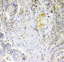
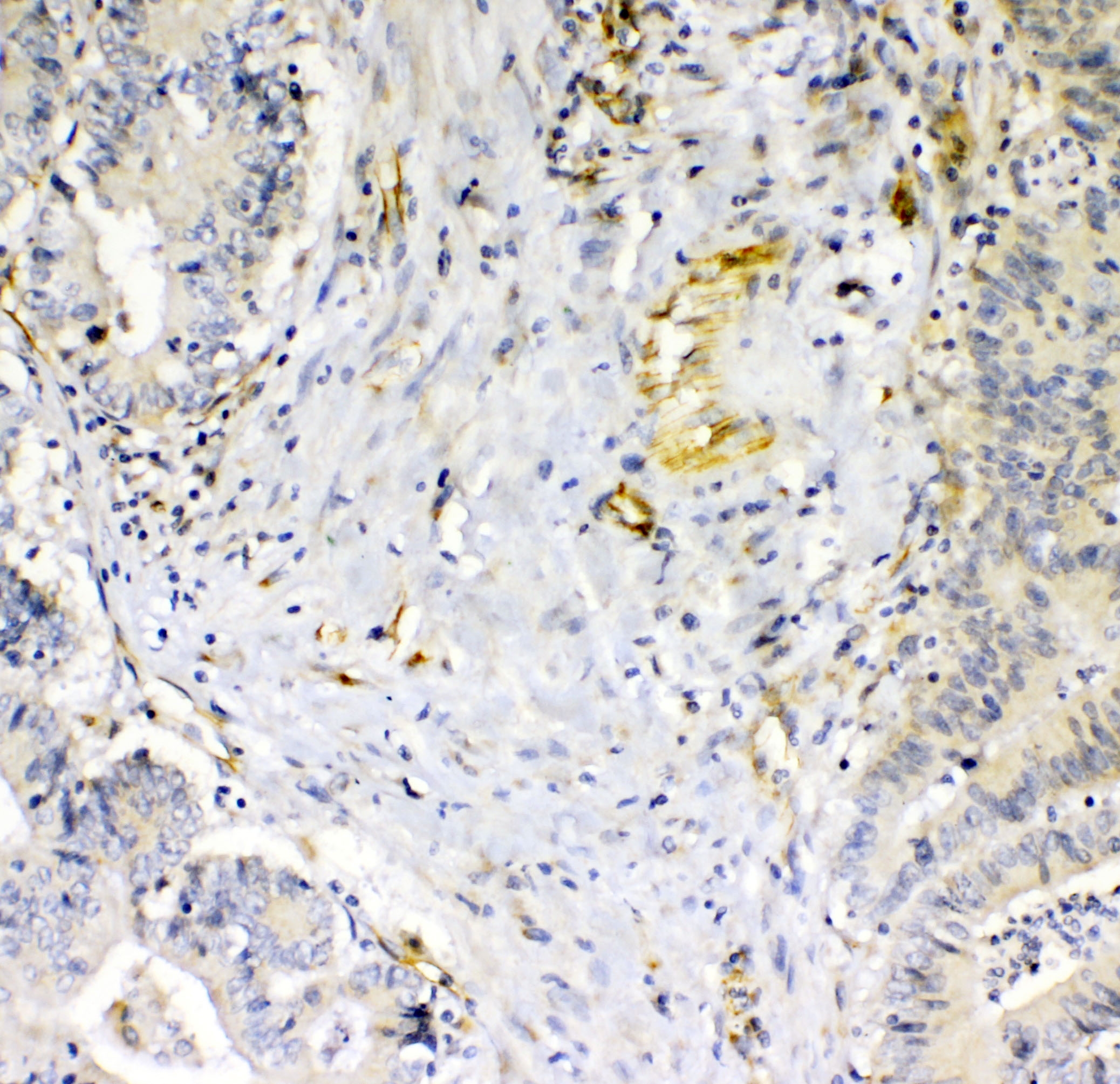
ARG40764 anti-VE Cadherin antibody IHC-P image
Immunohistochemistry: Paraffin-embedded Human mammary cancer tissue. Antigen Retrieval: Heat mediation was performed in Citrate buffer (pH 6.0, epitope retrieval solution) for 20 min. The tissue section was blocked with 10% goat serum. The tissue section was then stained with ARG40764 anti-VE Cadherin antibody at 1 µg/ml, overnight at 4°C.
-

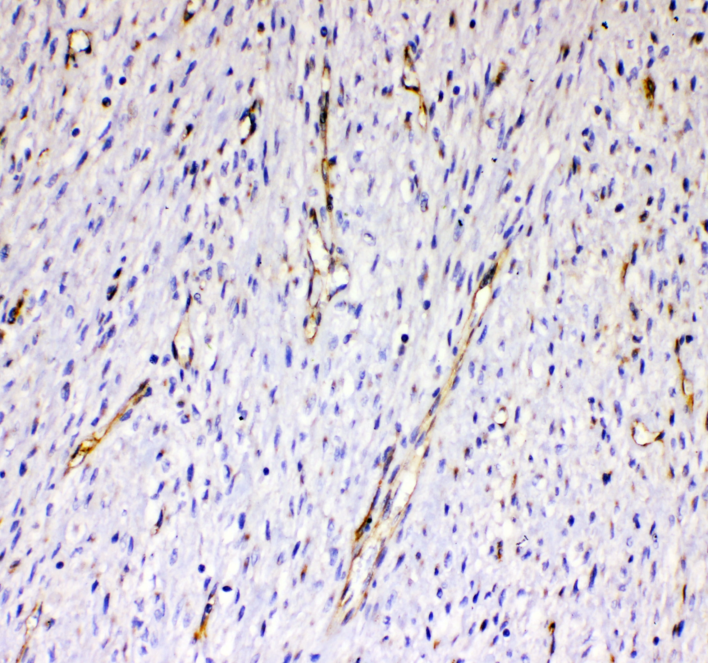
ARG40764 anti-VE Cadherin antibody IHC-P image
Immunohistochemistry: Paraffin-embedded Human mammary cancer tissue. Antigen Retrieval: Heat mediation was performed in Citrate buffer (pH 6.0, epitope retrieval solution) for 20 min. The tissue section was blocked with 10% goat serum. The tissue section was then stained with ARG40764 anti-VE Cadherin antibody at 1 µg/ml, overnight at 4°C.
-
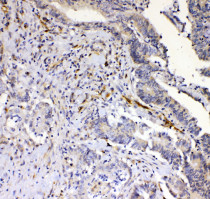
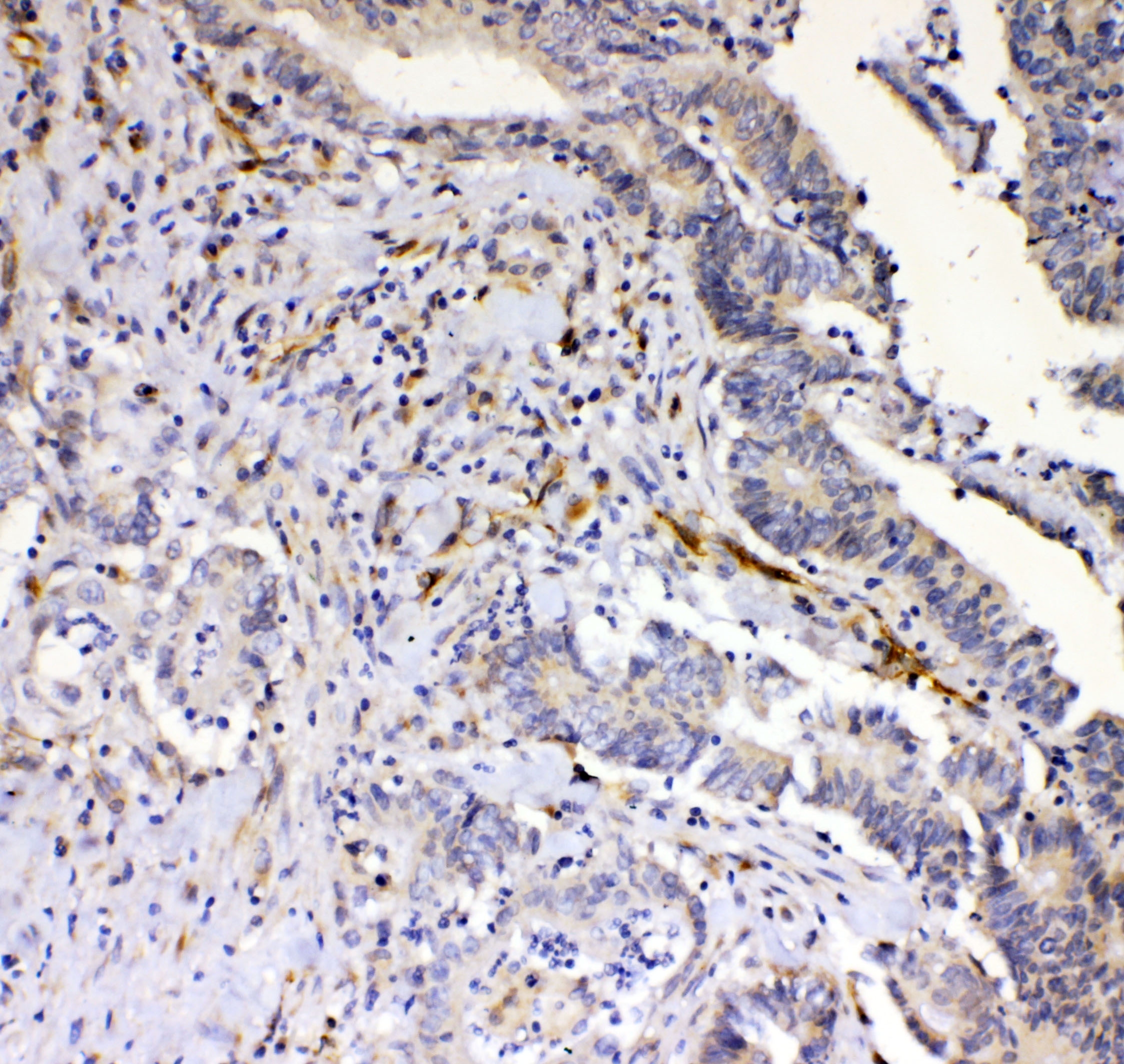
ARG40764 anti-VE Cadherin antibody IHC-P image
Immunohistochemistry: Paraffin-embedded Human rectal cancer tissue. Antigen Retrieval: Heat mediation was performed in Citrate buffer (pH 6.0, epitope retrieval solution) for 20 min. The tissue section was blocked with 10% goat serum. The tissue section was then stained with ARG40764 anti-VE Cadherin antibody at 1 µg/ml, overnight at 4°C.
-
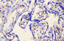
ARG40764 anti-VE Cadherin antibody IHC-Fr image
Immunohistochemistry: Frozen section of Human placenta tissue. The tissue section was blocked with 10% goat serum and then stained with ARG40764 anti-VE Cadherin antibody at 1 μg/ml dilution, overnight at 4°C.
-
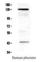
ARG40764 anti-VE Cadherin antibody WB image
Western blot: 50 µg of sample under reducing conditions. Human placenta tissue lysate stained with ARG40764 anti-VE Cadherin antibody at 0.5 µg/ml, overnight at 4°C.