ARG43066
anti-U2AF2 / U2AF65 antibody
anti-U2AF2 / U2AF65 antibody for Flow cytometry,ICC/IF,IHC-Formalin-fixed paraffin-embedded sections,Western blot and Human,Mouse,Rat
概述
| 产品描述 | Rabbit Polyclonal antibody recognizes U2AF2 / U2AF65 |
|---|---|
| 反应物种 | Hu, Ms, Rat |
| 应用 | FACS, ICC/IF, IHC-P, WB |
| 宿主 | Rabbit |
| 克隆 | Polyclonal |
| 同位型 | IgG |
| 靶点名称 | U2AF2 / U2AF65 |
| 抗原物种 | Human |
| 抗原 | Recombinant protein corresponding to M238-H470 of Human U2AF2 / U2AF65. |
| 偶联标记 | Un-conjugated |
| 別名 | U2 auxiliary factor 65 kDa subunit; U2AF65; U2 snRNP auxiliary factor large subunit; Splicing factor U2AF 65 kDa subunit; 65; hU2AF; hU2AF65 |
应用说明
| 应用建议 |
|
||||||||||
|---|---|---|---|---|---|---|---|---|---|---|---|
| 应用说明 | IHC-P: Antigen Retrieval: Heat mediation was performed in EDTA buffer (pH 8.0). * The dilutions indicate recommended starting dilutions and the optimal dilutions or concentrations should be determined by the scientist. |
属性
| 形式 | Liquid |
|---|---|
| 纯化 | Affinity purification with immunogen. |
| 缓冲液 | 0.2% Na2HPO4, 0.9% NaCl, 0.05% Sodium azide and 4% Trehalose. |
| 抗菌剂 | 0.05% Sodium azide |
| 稳定剂 | 4% Trehalose |
| 浓度 | 0.5 mg/ml |
| 存放说明 | For continuous use, store undiluted antibody at 2-8°C for up to a week. For long-term storage, aliquot and store at -20°C or below. Storage in frost free freezers is not recommended. Avoid repeated freeze/thaw cycles. Suggest spin the vial prior to opening. The antibody solution should be gently mixed before use. |
| 注意事项 | For laboratory research only, not for drug, diagnostic or other use. |
生物信息
| 数据库连接 |
Swiss-port # P26368 Human Splicing factor U2AF 65 kDa subunit Swiss-port # P26369 Mouse Splicing factor U2AF 65 kDa subunit |
|---|---|
| 基因名称 | U2AF2 |
| 全名 | U2 small nuclear RNA auxiliary factor 2 |
| 背景介绍 | U2 auxiliary factor (U2AF), comprised of a large and a small subunit, is a non-snRNP protein required for the binding of U2 snRNP to the pre-mRNA branch site. This gene encodes the U2AF large subunit which contains a sequence-specific RNA-binding region with 3 RNA recognition motifs and an Arg/Ser-rich domain necessary for splicing. The large subunit binds to the polypyrimidine tract of introns early during spliceosome assembly. Multiple transcript variants have been detected for this gene, but the full-length natures of only two have been determined to date. [provided by RefSeq, Jul 2008] |
| 生物功能 | Plays a role in pre-mRNA splicing and 3'-end processing (PubMed:17024186). By recruiting PRPF19 and the PRP19C/Prp19 complex/NTC/Nineteen complex to the RNA polymerase II C-terminal domain (CTD), and thereby pre-mRNA, may couple transcription to splicing (PubMed:21536736). Induces cardiac troponin-T (TNNT2) pre-mRNA exon inclusion in muscle. Regulates the TNNT2 exon 5 inclusion through competition with MBNL1. Binds preferentially to a single-stranded structure within the polypyrimidine tract of TNNT2 intron 4 during spliceosome assembly. Required for the export of mRNA out of the nucleus, even if the mRNA is encoded by an intron-less gene. Represses the splicing of MAPT/Tau exon 10. Positively regulates pre-mRNA 3'-end processing by recruiting the CFIm complex to cleavage and polyadenylation signals (PubMed:17024186). [UniProt] |
| 细胞定位 | Nucleus. [UniProt] |
| 预测分子量 | 54 kDa |
| 翻译后修饰 | Lysyl-hydroxylation at Lys-15 and Lys-276 affects the mRNA splicing activity of the protein, leading to regulate some, but not all, alternative splicing events. [UniProt] |
检测图片 (8) Click the Picture to Zoom In
-
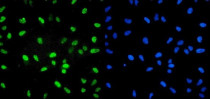
ARG43066 anti-U2AF2 / U2AF65 antibody ICC/IF image
Immunofluorescence: HeLa cells were blocked with 10% goat serum and then stained with ARG43066 anti-U2AF2 / U2AF65 antibody (green) at 2 µg/ml dilution, overnight at 4°C. DAPI (blue) for nuclear staining.
-

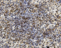
ARG43066 anti-U2AF2 / U2AF65 antibody IHC-P image
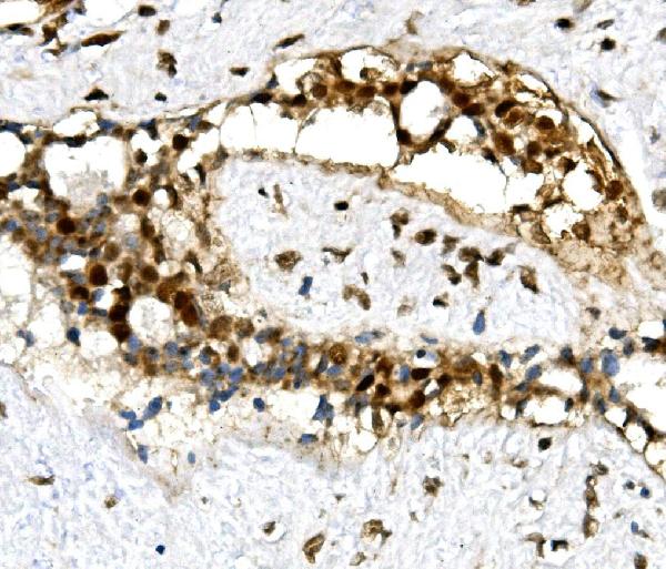
Immunohistochemistry: Paraffin-embedded Human mammary cancer tissue. Antigen Retrieval: Heat mediation was performed in EDTA buffer (pH 8.0). The tissue section was blocked with 10% goat serum. The tissue section was then stained with ARG43066 anti-U2AF2 / U2AF65 antibody at 1 µg/ml dilution, overnight at 4°C.
-

ARG43066 anti-U2AF2 / U2AF65 antibody WB image
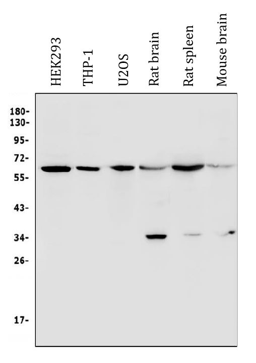
Western blot: 50 µg of sample under reducing conditions. HEK293, THP-1, U2OS, Rat brain, Rat spleen and Mouse brain lysates stained with ARG43066 anti-U2AF2 / U2AF65 antibody at 0.5 µg/ml dilution, overnight at 4°C.
-
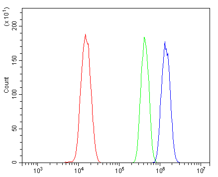
ARG43066 anti-U2AF2 / U2AF65 antibody FACS image
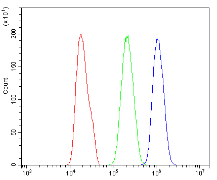
Flow Cytometry: PC-3 cells were blocked with 10% normal goat serum and then stained with ARG43066 anti-U2AF2 / U2AF65 antibody (blue) at 1 µg/10^6 cells for 30 min at 20°C, followed by incubation with DyLight®488 labelled secondary antibody. Isotype control antibody (green) was rabbit IgG (1 µg/10^6 cells) used under the same conditions. Unlabelled sample (red) was also used as a control.
-
ARG43066 anti-U2AF2 / U2AF65 antibody IHC-P image
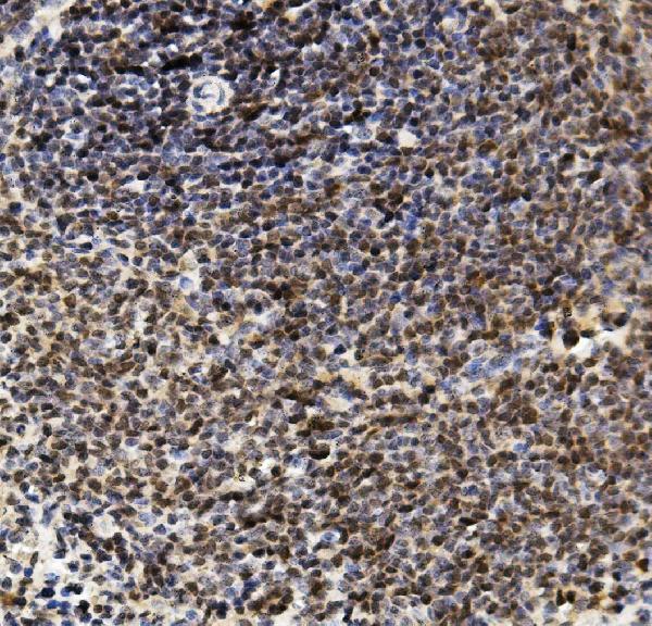
Immunohistochemistry: Paraffin-embedded Mouse spleen tissue. Antigen Retrieval: Heat mediation was performed in EDTA buffer (pH 8.0). The tissue section was blocked with 10% goat serum. The tissue section was then stained with ARG43066 anti-U2AF2 / U2AF65 antibody at 1 µg/ml dilution, overnight at 4°C.
-
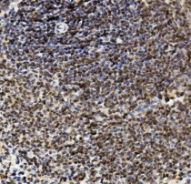
ARG43066 anti-U2AF2 / U2AF65 antibody IHC-P image
Immunohistochemistry: Paraffin-embedded Rat spleen tissue. Antigen Retrieval: Heat mediation was performed in EDTA buffer (pH 8.0). The tissue section was blocked with 10% goat serum. The tissue section was then stained with ARG43066 anti-U2AF2 / U2AF65 antibody at 1 µg/ml dilution, overnight at 4°C.
-

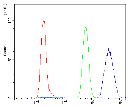
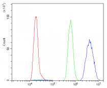
ARG43066 anti-U2AF2 / U2AF65 antibody FACS image
Flow Cytometry: ANA-1 cells were blocked with 10% normal goat serum and then stained with ARG43066 anti-U2AF2 / U2AF65 antibody (blue) at 1 µg/10^6 cells for 30 min at 20°C, followed by incubation with DyLight®488 labelled secondary antibody. Isotype control antibody (green) was rabbit IgG (1 µg/10^6 cells) used under the same conditions. Unlabelled sample (red) was also used as a control.
-
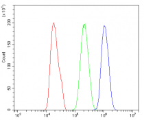
ARG43066 anti-U2AF2 / U2AF65 antibody FACS image
Flow Cytometry: NRK cells were blocked with 10% normal goat serum and then stained with ARG43066 anti-U2AF2 / U2AF65 antibody (blue) at 1 µg/10^6 cells for 30 min at 20°C, followed by incubation with DyLight®488 labelled secondary antibody. Isotype control antibody (green) was rabbit IgG (1 µg/10^6 cells) used under the same conditions. Unlabelled sample (red) was also used as a control.