ARG43095
anti-SRCIN1 / p140Cap antibody
anti-SRCIN1 / p140Cap antibody for Flow cytometry,IHC-Formalin-fixed paraffin-embedded sections,Western blot and Human,Mouse,Rat
概述
| 产品描述 | Rabbit Polyclonal antibody recognizes SRCIN1 / p140Cap |
|---|---|
| 反应物种 | Hu, Ms, Rat |
| 应用 | FACS, IHC-P, WB |
| 宿主 | Rabbit |
| 克隆 | Polyclonal |
| 同位型 | IgG |
| 靶点名称 | SRCIN1 / p140Cap |
| 抗原物种 | Human |
| 抗原 | Recombinant protein corresponding to E189-E287 of Human SRCIN1 / p140Cap. |
| 偶联标记 | Un-conjugated |
| 別名 | p130Cas-associated protein; SRC kinase signaling inhibitor 1; SNAP-25-interacting protein; p140Cap; SNIP; P140 |
应用说明
| 应用建议 |
|
||||||||
|---|---|---|---|---|---|---|---|---|---|
| 应用说明 | IHC-P: Antigen Retrieval: Heat mediation was performed in Citrate buffer (pH 6.0) for 20 min. * The dilutions indicate recommended starting dilutions and the optimal dilutions or concentrations should be determined by the scientist. |
||||||||
| 实际分子量 | ~ 140 kDa |
属性
| 形式 | Liquid |
|---|---|
| 纯化 | Affinity purification with immunogen. |
| 缓冲液 | 0.2% Na2HPO4, 0.9% NaCl, 0.05% Sodium azide and 4% Trehalose. |
| 抗菌剂 | 0.05% Sodium azide |
| 稳定剂 | 4% Trehalose |
| 浓度 | 0.5 mg/ml |
| 存放说明 | For continuous use, store undiluted antibody at 2-8°C for up to a week. For long-term storage, aliquot and store at -20°C or below. Storage in frost free freezers is not recommended. Avoid repeated freeze/thaw cycles. Suggest spin the vial prior to opening. The antibody solution should be gently mixed before use. |
| 注意事项 | For laboratory research only, not for drug, diagnostic or other use. |
生物信息
| 数据库连接 | |
|---|---|
| 基因名称 | SRCIN1 |
| 全名 | SRC kinase signaling inhibitor 1 |
| 生物功能 | Acts as a negative regulator of SRC by activating CSK which inhibits SRC activity and downstream signaling, leading to impaired cell spreading and migration. Regulates dendritic spine morphology. Involved in calcium-dependent exocytosis. May play a role in neurotransmitter release or synapse maintenance. [UniProt] |
| 细胞定位 | Cytoplasm, cytoskeleton. Cell projection, axon, dendrite. Cell junction, synapse, postsynaptic cell membrane, postsynaptic density. Note=Localized to the perinuclear region, lamellopodia, cortical actin and actin stress fibers but not to focal adhesions. Strongly expressed in axons and dendrites of the CA1 and CA3 hippocampal regions and of the dentate gyrus. Detected in both presynapses and postsynapses and in postsynaptic density fractions. [UniProt] |
| 预测分子量 | 127 kDa |
| 翻译后修饰 | Tyrosine-phosphorylated in response to EGF and to cell adhesion to integrin ligands. [UniProt] |
检测图片 (11) Click the Picture to Zoom In
-
ARG43095 anti-SRCIN1 / p140Cap antibody IHC-P image
Immunohistochemistry: Paraffin-embedded Human appendicitis tissue. Antigen Retrieval: Heat mediation was performed in Citrate buffer (pH 6.0) for 20 min. The tissue section was blocked with 10% goat serum. The tissue section was then stained with ARG43095 anti-SRCIN1 / p140Cap antibody at 1 µg/ml dilution, overnight at 4°C.
-
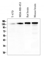
ARG43095 anti-SRCIN1 / p140Cap antibody WB image
Western blot: 50 µg of sample under reducing conditions. T-47D, MDA-MB-453, Rat brain and Mouse brain lysates stained with ARG43095 anti-SRCIN1 / p140Cap antibody at 0.5 µg/ml dilution, overnight at 4°C.
-
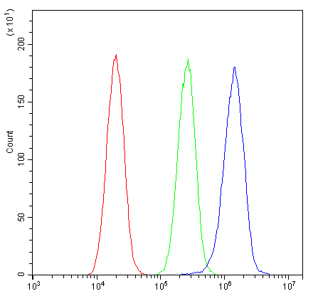
ARG43095 anti-SRCIN1 / p140Cap antibody FACS image
Flow Cytometry: U2OS cells were blocked with 10% normal goat serum and then stained with ARG43095 anti-SRCIN1 / p140Cap antibody (blue) at 1 µg/10^6 cells for 30 min at 20°C, followed by incubation with DyLight®488 labelled secondary antibody. Isotype control antibody (green) was rabbit IgG (1 µg/10^6 cells) used under the same conditions. Unlabelled sample (red) was also used as a control.
-
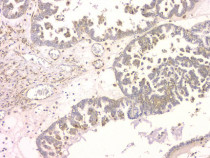
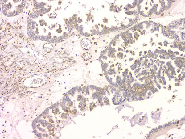
ARG43095 anti-SRCIN1 / p140Cap antibody IHC-P image
Immunohistochemistry: Paraffin-embedded Human ovary cancer tissue. Antigen Retrieval: Heat mediation was performed in Citrate buffer (pH 6.0) for 20 min. The tissue section was blocked with 10% goat serum. The tissue section was then stained with ARG43095 anti-SRCIN1 / p140Cap antibody at 1 µg/ml dilution, overnight at 4°C.
-
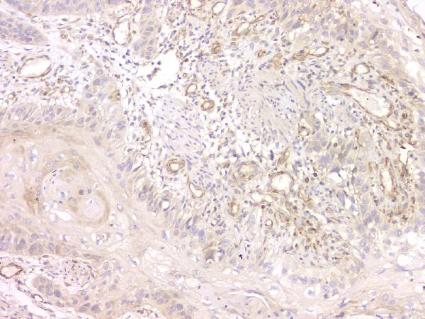
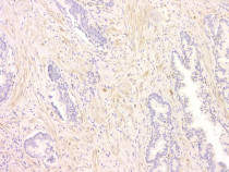
ARG43095 anti-SRCIN1 / p140Cap antibody IHC-P image
Immunohistochemistry: Paraffin-embedded Human oesophagus squama cancer tissue. Antigen Retrieval: Heat mediation was performed in Citrate buffer (pH 6.0) for 20 min. The tissue section was blocked with 10% goat serum. The tissue section was then stained with ARG43095 anti-SRCIN1 / p140Cap antibody at 1 µg/ml dilution, overnight at 4°C.
-
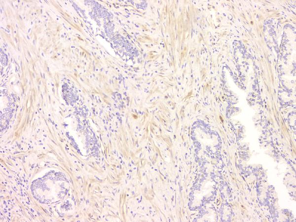
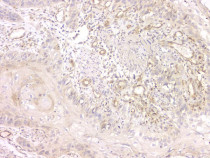
ARG43095 anti-SRCIN1 / p140Cap antibody IHC-P image
Immunohistochemistry: Paraffin-embedded Human ovary cancer tissue. Antigen Retrieval: Heat mediation was performed in Citrate buffer (pH 6.0) for 20 min. The tissue section was blocked with 10% goat serum. The tissue section was then stained with ARG43095 anti-SRCIN1 / p140Cap antibody at 1 µg/ml dilution, overnight at 4°C.
-
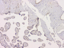
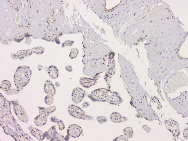
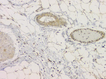
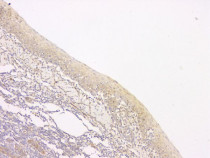
ARG43095 anti-SRCIN1 / p140Cap antibody IHC-P image
Immunohistochemistry: Paraffin-embedded Human placenta tissue. Antigen Retrieval: Heat mediation was performed in Citrate buffer (pH 6.0) for 20 min. The tissue section was blocked with 10% goat serum. The tissue section was then stained with ARG43095 anti-SRCIN1 / p140Cap antibody at 1 µg/ml dilution, overnight at 4°C.
-

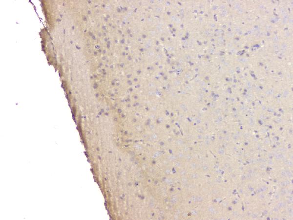
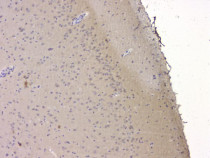
ARG43095 anti-SRCIN1 / p140Cap antibody IHC-P image
Immunohistochemistry: Paraffin-embedded Mouse brain tissue. Antigen Retrieval: Heat mediation was performed in Citrate buffer (pH 6.0) for 20 min. The tissue section was blocked with 10% goat serum. The tissue section was then stained with ARG43095 anti-SRCIN1 / p140Cap antibody at 1 µg/ml dilution, overnight at 4°C.
-
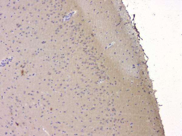
ARG43095 anti-SRCIN1 / p140Cap antibody IHC-P image
Immunohistochemistry: Paraffin-embedded Rat brain tissue. Antigen Retrieval: Heat mediation was performed in Citrate buffer (pH 6.0) for 20 min. The tissue section was blocked with 10% goat serum. The tissue section was then stained with ARG43095 anti-SRCIN1 / p140Cap antibody at 1 µg/ml dilution, overnight at 4°C.
-
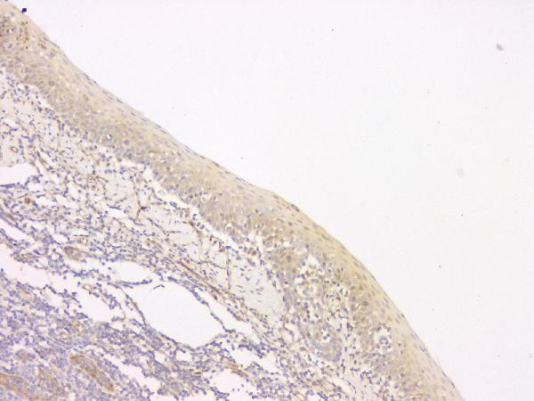
ARG43095 anti-SRCIN1 / p140Cap antibody IHC-P image
Immunohistochemistry: Paraffin-embedded Human tonsil tissue. Antigen Retrieval: Heat mediation was performed in Citrate buffer (pH 6.0) for 20 min. The tissue section was blocked with 10% goat serum. The tissue section was then stained with ARG43095 anti-SRCIN1 / p140Cap antibody at 1 µg/ml dilution, overnight at 4°C.
-

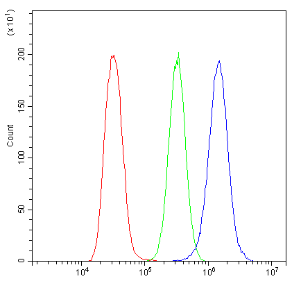
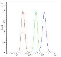
ARG43095 anti-SRCIN1 / p140Cap antibody FACS image
Flow Cytometry: A549 cells were blocked with 10% normal goat serum and then stained with ARG43095 anti-SRCIN1 / p140Cap antibody (blue) at 1 µg/10^6 cells for 30 min at 20°C, followed by incubation with DyLight®488 labelled secondary antibody. Isotype control antibody (green) was rabbit IgG (1 µg/10^6 cells) used under the same conditions. Unlabelled sample (red) was also used as a control.