ARG65163
anti-PDE1A antibody
anti-PDE1A antibody for IHC-Formalin-fixed paraffin-embedded sections,Western blot and Human
Signaling Transduction antibody
概述
| 产品描述 | Goat Polyclonal antibody recognizes PDE1A |
|---|---|
| 反应物种 | Hu |
| 预测物种 | Ms, Rat, Cow |
| 应用 | IHC-P, WB |
| 特异性 | This antibody is expected to recognize isoform 1 (NP_005010.2) only. |
| 宿主 | Goat |
| 克隆 | Polyclonal |
| 同位型 | IgG |
| 靶点名称 | PDE1A |
| 抗原物种 | Human |
| 抗原 | C-DLHKNSEDLVNAE |
| 偶联标记 | Un-conjugated |
| 別名 | CAM-PDE-1A; Cam-PDE 1A; HCAM1; Calcium/calmodulin-dependent 3',5'-cyclic nucleotide phosphodiesterase 1A; EC 3.1.4.17; HSPDE1A; hCam-1; HCAM-1; 61 kDa Cam-PDE |
应用说明
| 应用建议 |
|
||||||
|---|---|---|---|---|---|---|---|
| 应用说明 | WB: Recommend incubate at RT for 1h. IHC-P: Antigen Retrieval: Steam tissue section in Citrate buffer (pH 6.0). * The dilutions indicate recommended starting dilutions and the optimal dilutions or concentrations should be determined by the scientist. |
属性
| 形式 | Liquid |
|---|---|
| 纯化 | Purified from goat serum by antigen affinity chromatography. |
| 缓冲液 | Tris saline (pH 7.3), 0.02% Sodium azide and 0.5% BSA. |
| 抗菌剂 | 0.02% Sodium azide |
| 稳定剂 | 0.5% BSA |
| 浓度 | 0.5 mg/ml |
| 存放说明 | For continuous use, store undiluted antibody at 2-8°C for up to a week. For long-term storage, aliquot and store at -20°C or below. Storage in frost free freezers is not recommended. Avoid repeated freeze/thaw cycles. Suggest spin the vial prior to opening. The antibody solution should be gently mixed before use. |
| 注意事项 | For laboratory research only, not for drug, diagnostic or other use. |
生物信息
| 数据库连接 |
Swiss-port # P54750 Human Calcium/calmodulin-dependent 3',5'-cyclic nucleotide phosphodiesterase 1A |
|---|---|
| 背景介绍 | Cyclic nucleotide phosphodiesterases (PDEs) play a role in signal transduction by regulating intracellular cyclic nucleotide concentrations through hydrolysis of cAMP and/or cGMP to their respective nucleoside 5-prime monophosphates. Members of the PDE1 family, such as PDE1A, are Ca(2+)/calmodulin (see CALM1; MIM 114180)-dependent PDEs (CaM-PDEs) that are activated by calmodulin in the presence of Ca(2+) (Michibata et al., 2001 [PubMed 11342109]; Fidock et al., 2002 [PubMed 11747989]).[supplied by OMIM, Oct 2009] |
| 研究领域 | Signaling Transduction antibody |
| 预测分子量 | 61 kDa |
检测图片 (2) Click the Picture to Zoom In
-
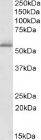
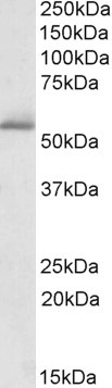
ARG65163 anti-PDE1A antibody WB image
Western Blot: Human Cerebellum lysate (35 µg protein in RIPA buffer) stained with ARG65163 anti-PDE1A antibody at 1 µg/ml dilution.
-
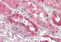
ARG65163 anti-PDE1A antibody IHC-P image
Immunohistochemistry: Paraffin-embedded Human kidney tissue. Antigen Retrieval: Steam tissue section in Citrate buffer (pH 6.0). The tissue section was stained with ARG65163 anti-PDE1A antibody at 3.75 µg/ml dilution followed by AP-staining.