ARG40514
anti-OAT antibody
anti-OAT antibody for Flow cytometry,ICC/IF,IHC-Formalin-fixed paraffin-embedded sections,Western blot and Human,Mouse,Rat
概述
| 产品描述 | Rabbit Polyclonal antibody recognizes OAT |
|---|---|
| 反应物种 | Hu, Ms, Rat |
| 应用 | FACS, ICC/IF, IHC-P, WB |
| 宿主 | Rabbit |
| 克隆 | Polyclonal |
| 同位型 | IgG |
| 靶点名称 | OAT |
| 抗原物种 | Human |
| 抗原 | KLH-conjugated synthetic peptide corresponding to aa. 27-55 of Human OAT. |
| 偶联标记 | Un-conjugated |
| 別名 | OATASE; OKT; Ornithine aminotransferase, mitochondrial; Ornithine--oxo-acid aminotransferase; GACR; HOGA; Ornithine delta-aminotransferase; EC 2.6.1.13 |
应用说明
| 应用建议 |
|
||||||||||
|---|---|---|---|---|---|---|---|---|---|---|---|
| 应用说明 | * The dilutions indicate recommended starting dilutions and the optimal dilutions or concentrations should be determined by the scientist. | ||||||||||
| 阳性对照 | 293 |
属性
| 形式 | Liquid |
|---|---|
| 纯化 | Purification with Protein A and immunogen peptide. |
| 缓冲液 | PBS and 0.09% (W/V) Sodium azide. |
| 抗菌剂 | 0.09% (W/V) Sodium azide. |
| 存放说明 | For continuous use, store undiluted antibody at 2-8°C for up to a week. For long-term storage, aliquot and store at -20°C or below. Storage in frost free freezers is not recommended. Avoid repeated freeze/thaw cycles. Suggest spin the vial prior to opening. The antibody solution should be gently mixed before use. |
| 注意事项 | For laboratory research only, not for drug, diagnostic or other use. |
生物信息
| 数据库连接 | |
|---|---|
| 基因名称 | OAT |
| 全名 | ornithine aminotransferase |
| 背景介绍 | This gene encodes the mitochondrial enzyme ornithine aminotransferase, which is a key enzyme in the pathway that converts arginine and ornithine into the major excitatory and inhibitory neurotransmitters glutamate and GABA. Mutations that result in a deficiency of this enzyme cause the autosomal recessive eye disease Gyrate Atrophy. Alternatively spliced transcript variants encoding different isoforms have been described. Related pseudogenes have been defined on the X chromosome. [provided by RefSeq, Jan 2010] |
| 细胞定位 | Mitochondrion matrix. [UniProt] |
| 预测分子量 | 49 kDa |
检测图片 (4) Click the Picture to Zoom In
-

ARG40514 anti-OAT antibody ICC/IF image
Immunofluorescence: A549 cells were fixed with 4% PFA (20 min), permeabilized with Triton X-100 (0.1%, 10 min), then stained with ARG40514 anti-OAT antibody (green) at 1:25, 1 hour at 37°C. Cytoplasmic actin was counterstained with Alexa Fluor® 555 conjugated Phalloidin (red).
-
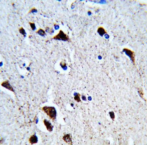
ARG40514 anti-OAT antibody IHC-P image
Immunohistochemistry: Formalin-fixed and paraffin-embedded Human brain tissue stained with ARG40514 anti-OAT antibody.
-

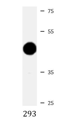
ARG40514 anti-OAT antibody WB image
Western blot: 20 µg of 293 whole cell lysate stained with ARG40514 anti-OAT antibody at 1:1000 dilution.
-

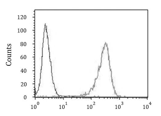
ARG40514 anti-OAT antibody FACS image
Flow Cytometry: 293 cells stained with ARG40514 anti-OAT antibody (right histogram) or without primary antibody as control (left histogram), followed by incubation with FITC labelled secondary antibody.