ARG40378
anti-MAOA antibody
anti-MAOA antibody for Flow cytometry,ICC/IF,IHC-Formalin-fixed paraffin-embedded sections,Western blot and Human,Mouse,Rat
概述
| 产品描述 | Rabbit Polyclonal antibody recognizes MAOA |
|---|---|
| 反应物种 | Hu, Ms, Rat |
| 应用 | FACS, ICC/IF, IHC-P, WB |
| 宿主 | Rabbit |
| 克隆 | Polyclonal |
| 同位型 | IgG |
| 靶点名称 | MAOA |
| 抗原物种 | Human |
| 抗原 | Synthetic peptide corresponding to aa. 457-493 of Human MAOA. (REVLNGLGKVTEKDIWVQEPESKDVPAVEITHTFWER) |
| 偶联标记 | Un-conjugated |
| 別名 | MAO-A; EC 1.4.3.4; Amine oxidase [flavin-containing] A; Monoamine oxidase type A |
应用说明
| 应用建议 |
|
||||||||||
|---|---|---|---|---|---|---|---|---|---|---|---|
| 应用说明 | IHC-P: Antigen Retrieval: By heat mediation. * The dilutions indicate recommended starting dilutions and the optimal dilutions or concentrations should be determined by the scientist. |
||||||||||
| 实际分子量 | 60 kDa |
属性
| 形式 | Liquid |
|---|---|
| 纯化 | Affinity purification with immunogen. |
| 缓冲液 | 0.2% Na2HPO4, 0.9% NaCl, 0.05% Sodium azide and 5% BSA. |
| 抗菌剂 | 0.05% Sodium azide |
| 稳定剂 | 5% BSA |
| 浓度 | 0.5 mg/ml |
| 存放说明 | For continuous use, store undiluted antibody at 2-8°C for up to a week. For long-term storage, aliquot and store at -20°C or below. Storage in frost free freezers is not recommended. Avoid repeated freeze/thaw cycles. Suggest spin the vial prior to opening. The antibody solution should be gently mixed before use. |
| 注意事项 | For laboratory research only, not for drug, diagnostic or other use. |
生物信息
| 数据库连接 |
Swiss-port # P21397 Human Amine oxidase [flavin-containing] A Swiss-port # Q64133 Mouse Amine oxidase [flavin-containing] A |
|---|---|
| 基因名称 | MAOA |
| 全名 | monoamine oxidase A |
| 背景介绍 | This gene is one of two neighboring gene family members that encode mitochondrial enzymes which catalyze the oxidative deamination of amines, such as dopamine, norepinephrine, and serotonin. Mutation of this gene results in Brunner syndrome. This gene has also been associated with a variety of other psychiatric disorders, including antisocial behavior. Alternatively spliced transcript variants encoding multiple isoforms have been observed. [provided by RefSeq, Jul 2012] |
| 生物功能 | Catalyzes the oxidative deamination of biogenic and xenobiotic amines and has important functions in the metabolism of neuroactive and vasoactive amines in the central nervous system and peripheral tissues. MAOA preferentially oxidizes biogenic amines such as 5-hydroxytryptamine (5-HT), norepinephrine and epinephrine. [UniProt] |
| 细胞定位 | Mitochondrion outer membrane; Single-pass type IV membrane protein; Cytoplasmic side. [UniProt] |
| 预测分子量 | 60 kDa |
检测图片 (7) Click the Picture to Zoom In
-
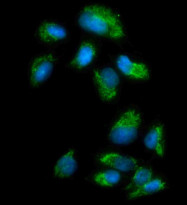
ARG40378 anti-MAOA antibody ICC/IF image
Immunofluorescence: U2OS cells were blocked with 10% goat serum and then stained with ARG40378 anti-MAOA antibody (green) at 5 µg/ml dilution, overnight at 4°C. DAPI (blue) for nuclear staining.
-
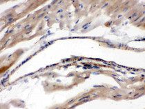
ARG40378 anti-MAOA antibody IHC-P image
Immunohistochemistry: Paraffin-embedded Mouse cardiac muscle stained with ARG40378 anti-MAOA antibody.
-
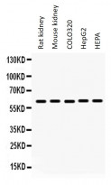
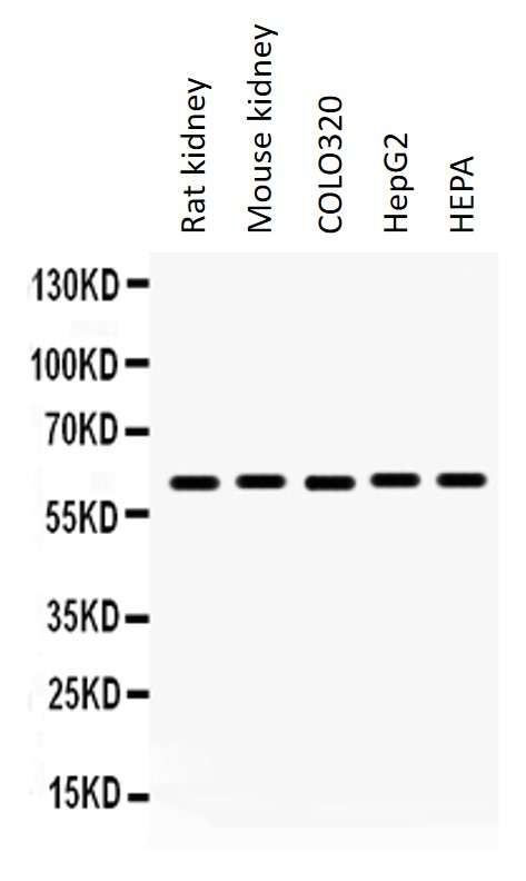
ARG40378 anti-MAOA antibody WB image
Western blot: 50 µg Rat kidney, 50 µg of Mouse kidney, 40 µg of COLO320, 40 µg of HepG2 and 40 µg of HEPA whole cell lysates stained with ARG40378 anti-MAOA antibody at 0.5 µg/ml dilution.
-
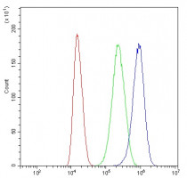
ARG40378 anti-MAOA antibody FACS image
Flow Cytometry: U87 cells were blocked with 10% normal goat serum and then stained with ARG40378 anti-MAOA antibody (blue) at 1 µg/10^6 cells for 30 min at 20°C, followed by incubation with DyLight®488 labelled secondary antibody. Isotype control antibody (green) was rabbit IgG (1 µg/10^6 cells) used under the same conditions. Unlabelled sample (red) was also used as a control.
-
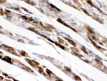
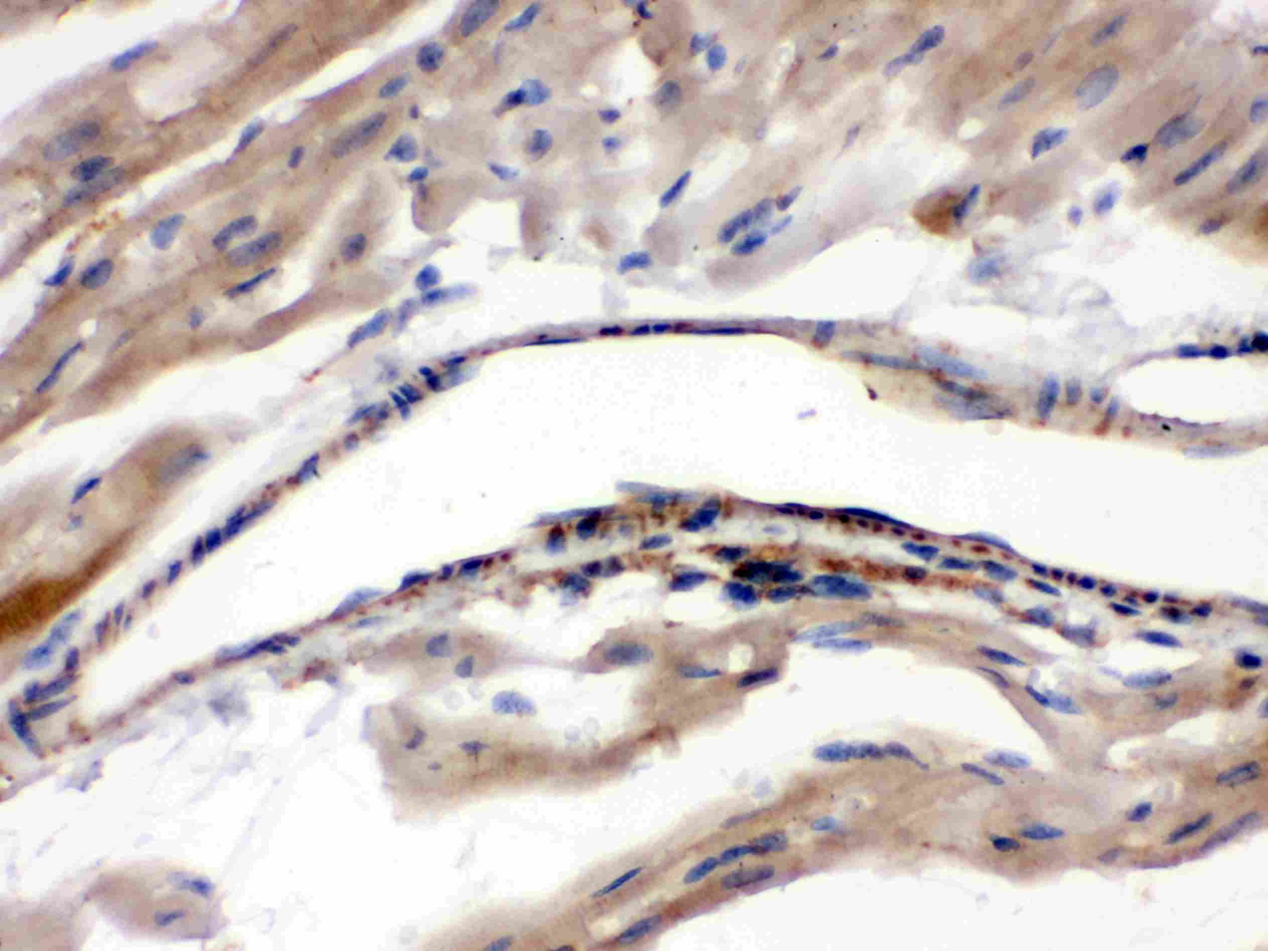
ARG40378 anti-MAOA antibody IHC-P image
Immunohistochemistry: Paraffin-embedded Rat cardiac muscle stained with ARG40378 anti-MAOA antibody.
-

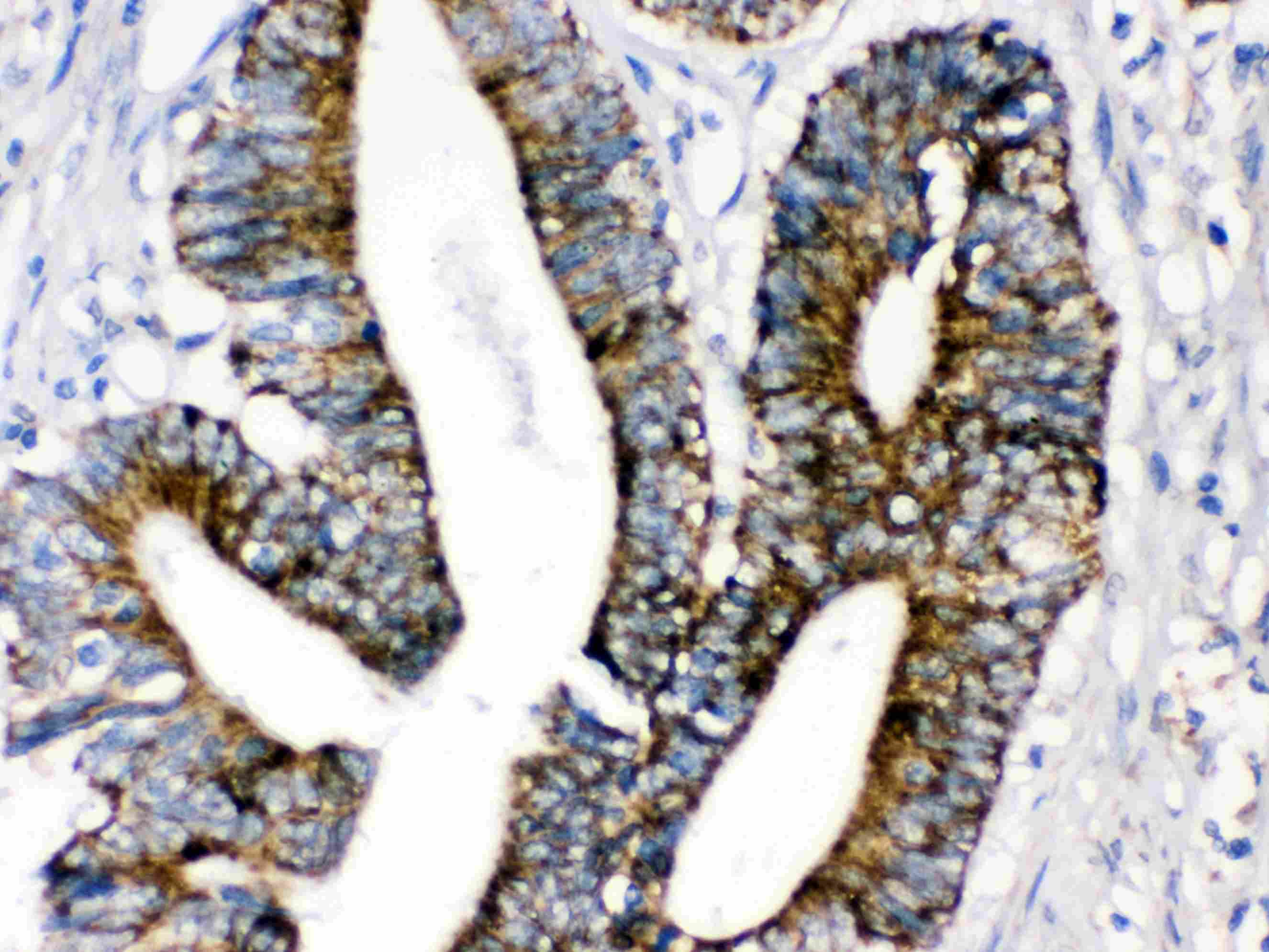
ARG40378 anti-MAOA antibody IHC-P image
Immunohistochemistry: Paraffin-embedded Human intestinal cancer tissue stained with ARG40378 anti-MAOA antibody.
-
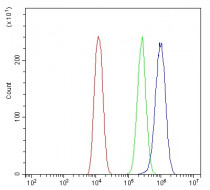
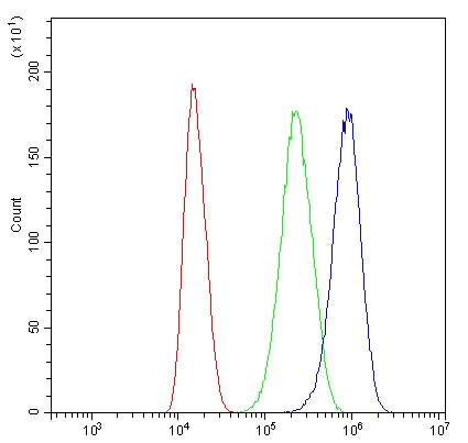
ARG40378 anti-MAOA antibody FACS image
Flow Cytometry: U2OS cells were blocked with 10% normal goat serum and then stained with ARG40378 anti-MAOA antibody (blue) at 1 µg/10^6 cells for 30 min at 20°C, followed by incubation with DyLight®488 labelled secondary antibody. Isotype control antibody (green) was rabbit IgG (1 µg/10^6 cells) used under the same conditions. Unlabelled sample (red) was also used as a control.