ARG10718
anti-Lamin A + C antibody
anti-Lamin A + C antibody for ICC/IF,IHC-Frozen sections,Western blot and Human,Mouse,Rat,Cow,Horse,Pig
概述
| 产品描述 | Rabbit Polyclonal antibody recognizes Lamin A + C |
|---|---|
| 反应物种 | Hu, Ms, Rat, Cow, Hrs, Pig |
| 预测物种 | Chk |
| 应用 | ICC/IF, IHC-Fr, WB |
| 宿主 | Rabbit |
| 克隆 | Polyclonal |
| 同位型 | IgG |
| 靶点名称 | Lamin A + C |
| 抗原物种 | Human |
| 抗原 | Full length recombinant Human Lamin A purified from E. coli. |
| 偶联标记 | Un-conjugated |
| 別名 | HGPS; Renal carcinoma antigen NY-REN-32; LDP1; FPL; LMN1; CDCD1; LMNL1; CDDC; PRO1; EMD2; CMT2B1; 70 kDa lamin; LFP; Prelamin-A/C; LMNC; FPLD2; LGMD1B; IDC; FPLD; CMD1A |
应用说明
| 应用建议 |
|
||||||||
|---|---|---|---|---|---|---|---|---|---|
| 应用说明 | * The dilutions indicate recommended starting dilutions and the optimal dilutions or concentrations should be determined by the scientist. |
属性
| 形式 | Liquid |
|---|---|
| 纯化 | Unpurified. |
| 缓冲液 | Serum. |
| 存放说明 | For continuous use, store undiluted antibody at 2-8°C for up to a week. For long-term storage, aliquot and store at -20°C or below. Storage in frost free freezers is not recommended. Avoid repeated freeze/thaw cycles. Suggest spin the vial prior to opening. The antibody solution should be gently mixed before use. |
| 注意事项 | For laboratory research only, not for drug, diagnostic or other use. |
生物信息
| 数据库连接 | |
|---|---|
| 基因名称 | LMNA |
| 全名 | lamin A/C |
| 背景介绍 | The nuclear lamina consists of a two-dimensional matrix of proteins located next to the inner nuclear membrane. The lamin family of proteins make up the matrix and are highly conserved in evolution. During mitosis, the lamina matrix is reversibly disassembled as the lamin proteins are phosphorylated. Lamin proteins are thought to be involved in nuclear stability, chromatin structure and gene expression. Vertebrate lamins consist of two types, A and B. Alternative splicing results in multiple transcript variants. Mutations in this gene lead to several diseases: Emery-Dreifuss muscular dystrophy, familial partial lipodystrophy, limb girdle muscular dystrophy, dilated cardiomyopathy, Charcot-Marie-Tooth disease, and Hutchinson-Gilford progeria syndrome. [provided by RefSeq, Apr 2012] |
| 生物功能 | Lamins are components of the nuclear lamina, a fibrous layer on the nucleoplasmic side of the inner nuclear membrane, which is thought to provide a framework for the nuclear envelope and may also interact with chromatin. Lamin A and C are present in equal amounts in the lamina of mammals. Plays an important role in nuclear assembly, chromatin organization, nuclear membrane and telomere dynamics. Required for normal development of peripheral nervous system and skeletal muscle and for muscle satellite cell proliferation. Required for osteoblastogenesis and bone formation. Also prevents fat infiltration of muscle and bone marrow, helping to maintain the volume and strength of skeletal muscle and bone. Prelamin-A/C can accelerate smooth muscle cell senescence. It acts to disrupt mitosis and induce DNA damage in vascular smooth muscle cells (VSMCs), leading to mitotic failure, genomic instability, and premature senescence. [UniProt] |
| 预测分子量 | Lamin A: 74 kDa Lamin C: 65 kDa |
| 翻译后修饰 | Increased phosphorylation of the lamins occurs before envelope disintegration and probably plays a role in regulating lamin associations. Proteolytic cleavage of the C-terminal of 18 residues of prelamin-A/C results in the production of lamin-A/C. The prelamin-A/C maturation pathway includes farnesylation of CAAX motif, ZMPSTE24/FACE1 mediated cleavage of the last three amino acids, methylation of the C-terminal cysteine and endoproteolytic removal of the last 15 C-terminal amino acids. Proteolytic cleavage requires prior farnesylation and methylation, and absence of these blocks cleavage. Sumoylation is necessary for the localization to the nuclear envelope. Farnesylation of prelamin-A/C facilitates nuclear envelope targeting. |
检测图片 (2) Click the Picture to Zoom In
-
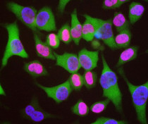
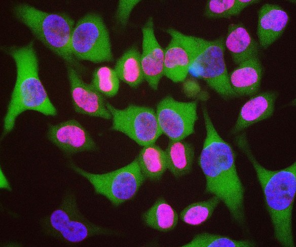
ARG10718 anti-Lamin A + C antibody ICC/IF image
Immunocytochemistry: HeLa cells stained with ARG10718 anti-Lamin A + C antibody (red) and co-stained with a monoclonal 6H11 to HSP27 (green) and DNA (blue). ARG10718 reveals strong nuclear lamina staining, while the Clone 6H11 reveals strong cytoplasmic staining. Since both DNA (blue) and Lamin A/C (red) are associated with the nuclear compartment, this region appears crimson in this image.
-

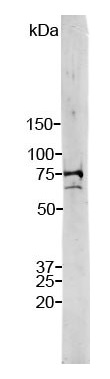
ARG10718 anti-Lamin A + C antibody WB image
Western blot: Strip crude HeLa cell extract stained with ARG10718 anti-Lamin A + C antibody. Note two strong and clean bands at 74 kDa and 65 kDa, corresponding to Lamin A and C.