ARG43051
anti-KIF15 antibody
anti-KIF15 antibody for Flow cytometry,ICC/IF,IHC-Formalin-fixed paraffin-embedded sections,Western blot and Human,Rat
概述
| 产品描述 | Rabbit Polyclonal antibody recognizes KIF15 |
|---|---|
| 反应物种 | Hu, Rat |
| 应用 | FACS, ICC/IF, IHC-P, WB |
| 宿主 | Rabbit |
| 克隆 | Polyclonal |
| 同位型 | IgG |
| 靶点名称 | KIF15 |
| 抗原物种 | Human |
| 抗原 | Recombinant protein corresponding to M1201-S1388 of Human KIF15. |
| 偶联标记 | Un-conjugated |
| 別名 | NY-BR-62; Kinesin-like protein 2; Serologically defined breast cancer antigen NY-BR-62; KNSL7; Kinesin-like protein KIF15; Kinesin-like protein 7; HKLP2; hKLP2 |
应用说明
| 应用建议 |
|
||||||||||
|---|---|---|---|---|---|---|---|---|---|---|---|
| 应用说明 | IHC-P: Antigen Retrieval: Heat mediation was performed in EDTA buffer (pH 8.0). * The dilutions indicate recommended starting dilutions and the optimal dilutions or concentrations should be determined by the scientist. |
属性
| 形式 | Liquid |
|---|---|
| 纯化 | Affinity purification with immunogen. |
| 缓冲液 | 0.2% Na2HPO4, 0.9% NaCl, 0.05% Sodium azide and 4% Trehalose. |
| 抗菌剂 | 0.05% Sodium azide |
| 稳定剂 | 4% Trehalose |
| 浓度 | 0.5 mg/ml |
| 存放说明 | For continuous use, store undiluted antibody at 2-8°C for up to a week. For long-term storage, aliquot and store at -20°C or below. Storage in frost free freezers is not recommended. Avoid repeated freeze/thaw cycles. Suggest spin the vial prior to opening. The antibody solution should be gently mixed before use. |
| 注意事项 | For laboratory research only, not for drug, diagnostic or other use. |
生物信息
| 数据库连接 | |
|---|---|
| 基因名称 | KIF15 |
| 全名 | kinesin family member 15 |
| 生物功能 | Plus-end directed kinesin-like motor enzyme involved in mitotic spindle assembly. [UniProt] |
| 细胞定位 | Cytoplasm. Cytoskeleton, spindle. Note=Detected during the interphase in the cytoplasm as finely punctuate pattern and irregularly shaped dots. Detected during mitosis on the mitotic spindle. Colocalizes with TPX2 in mitosis. Localizes at the central spindle at anaphase. Localizes at the sites of invaginating cell membranes, a position that corresponds to the location of the contractile actomyosin ring of dividing cells. Colocalizes with actin in interphase. [UniProt] |
| 预测分子量 | 160 kDa |
检测图片 (6) Click the Picture to Zoom In
-
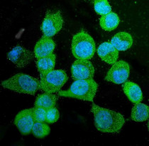
ARG43051 anti-KIF15 antibody ICC/IF image
Immunofluorescence: A431 cells were blocked with 10% goat serum and then stained with ARG43051 anti-KIF15 antibody (green) at 5 µg/ml dilution, overnight at 4°C. DAPI (blue) for nuclear staining.
-

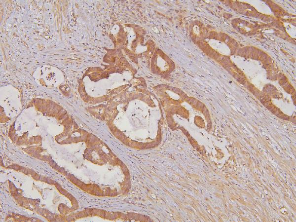
ARG43051 anti-KIF15 antibody IHC-P image
Immunohistochemistry: Paraffin-embedded Human colon cancer tissue. Antigen Retrieval: Heat mediation was performed in EDTA buffer (pH 8.0). The tissue section was blocked with 10% goat serum. The tissue section was then stained with ARG43051 anti-KIF15 antibody at 1 µg/ml dilution, overnight at 4°C.
-
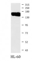
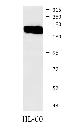
ARG43051 anti-KIF15 antibody WB image
Western blot: 50 µg of sample under reducing conditions. HL-60 whole cell lysate stained with ARG43051 anti-KIF15 antibody at 0.5 µg/ml dilution, overnight at 4°C.
-
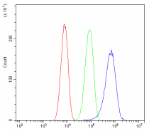
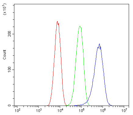
ARG43051 anti-KIF15 antibody FACS image
Flow Cytometry: SiHa cells were blocked with 10% normal goat serum and then stained with ARG43051 anti-KIF15 antibody (blue) at 1 µg/10^6 cells for 30 min at 20°C, followed by incubation with DyLight®488 labelled secondary antibody. Isotype control antibody (green) was rabbit IgG (1 µg/10^6 cells) used under the same conditions. Unlabelled sample (red) was also used as a control.
-
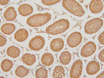
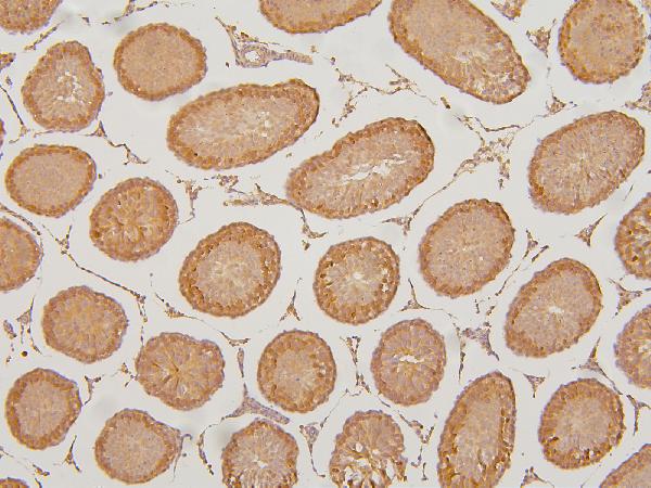
ARG43051 anti-KIF15 antibody IHC-P image
Immunohistochemistry: Paraffin-embedded Rat testis tissue. Antigen Retrieval: Heat mediation was performed in EDTA buffer (pH 8.0). The tissue section was blocked with 10% goat serum. The tissue section was then stained with ARG43051 anti-KIF15 antibody at 1 µg/ml dilution, overnight at 4°C.
-
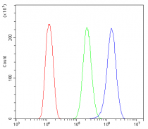
ARG43051 anti-KIF15 antibody FACS image
Flow Cytometry: U2OS cells were blocked with 10% normal goat serum and then stained with ARG43051 anti-KIF15 antibody (blue) at 1 µg/10^6 cells for 30 min at 20°C, followed by incubation with DyLight®488 labelled secondary antibody. Isotype control antibody (green) was rabbit IgG (1 µg/10^6 cells) used under the same conditions. Unlabelled sample (red) was also used as a control.