ARG42022
anti-KEAP1 antibody
anti-KEAP1 antibody for ICC/IF,IHC-Formalin-fixed paraffin-embedded sections,Western blot and Human,Mouse
概述
| 产品描述 | Goat Polyclonal antibody recognizes KEAP1 |
|---|---|
| 反应物种 | Hu, Ms |
| 预测物种 | Cow, Rat, Dog, Pig |
| 应用 | ICC/IF, IHC-P, WB |
| 宿主 | Goat |
| 克隆 | Polyclonal |
| 同位型 | IgG |
| 靶点名称 | KEAP1 |
| 抗原物种 | Human |
| 抗原 | Synthetic peptide around the internal region of Human KEAP1. (NP_036421.2) (C-EVTPSQHGNRTFS) |
| 偶联标记 | Un-conjugated |
| 別名 | KLHL19; Cytosolic inhibitor of Nrf2; INrf2; Kelch-like protein 19; Kelch-like ECH-associated protein 1 |
应用说明
| 应用建议 |
|
||||||||
|---|---|---|---|---|---|---|---|---|---|
| 应用说明 | IHC-P: Antigen Retrieval: Steam tissue section in Citrate buffer (pH 6.0). WB: Recommend incubate at RT for 1h. * The dilutions indicate recommended starting dilutions and the optimal dilutions or concentrations should be determined by the scientist. |
||||||||
| 阳性对照 | NIH/3T3 | ||||||||
| 实际分子量 | ~ 75 kDa |
属性
| 形式 | Liquid |
|---|---|
| 纯化 | Affinity purified |
| 缓冲液 | Tris saline (pH 7.3), 0.02% Sodium azide and 0.5% BSA. |
| 抗菌剂 | 0.02% Sodium azide |
| 稳定剂 | 0.5% BSA |
| 浓度 | 0.5 mg/ml |
| 存放说明 | For continuous use, store undiluted antibody at 2-8°C for up to a week. For long-term storage, aliquot and store at -20°C or below. Storage in frost free freezers is not recommended. Avoid repeated freeze/thaw cycles. Suggest spin the vial prior to opening. The antibody solution should be gently mixed before use. |
| 注意事项 | For laboratory research only, not for drug, diagnostic or other use. |
生物信息
| 数据库连接 |
Swiss-port # Q14145 Human Kelch-like ECH-associated protein 1 Swiss-port # Q9Z2X8 Mouse Kelch-like ECH-associated protein 1 |
|---|---|
| 基因名称 | KEAP1 |
| 全名 | kelch-like ECH-associated protein 1 |
| 背景介绍 | This gene encodes a protein containing KELCH-1 like domains, as well as a BTB/POZ domain. Kelch-like ECH-associated protein 1 interacts with NF-E2-related factor 2 in a redox-sensitive manner and the dissociation of the proteins in the cytoplasm is followed by transportation of NF-E2-related factor 2 to the nucleus. This interaction results in the expression of the catalytic subunit of gamma-glutamylcysteine synthetase. Two alternatively spliced transcript variants encoding the same isoform have been found for this gene. [provided by RefSeq, Jul 2008] |
| 生物功能 | Acts as a substrate adapter protein for the E3 ubiquitin ligase complex formed by CUL3 and RBX1 and targets NFE2L2/NRF2 for ubiquitination and degradation by the proteasome, thus resulting in the suppression of its transcriptional activity and the repression of antioxidant response element-mediated detoxifying enzyme gene expression. Retains NFE2L2/NRF2 and may also retain BPTF in the cytosol. Targets PGAM5 for ubiquitination and degradation by the proteasome. [UniProt] |
| 细胞定位 | Cytoplasm. Nucleus. Note=Shuttles between cytoplasm and nucleus. [UniProt] |
| 产品亮点 | Related products: KEAP1 antibodies; KEAP1 Duos / Panels; Anti-Goat IgG secondary antibodies; Related news: Keap1-Nrf2-ARE antibody panel is launched |
| 预测分子量 | 70 kDa |
| 翻译后修饰 | Ubiquitinated by the E3 ubiquitin ligase complex formed by CUL3 and RBX1 and is subject to proteasomal-independent degradation. Quinone-induced oxidative stress, but not sulforaphane, increases its ubiquitination. Ubiquitination and subsequent degradation is most pronounced following prolonged exposure of cells to oxidative stress, particularly in glutathione-deficient cells that are highly susceptible to oxidative stress. [UniProt] |
检测图片 (3) Click the Picture to Zoom In
-

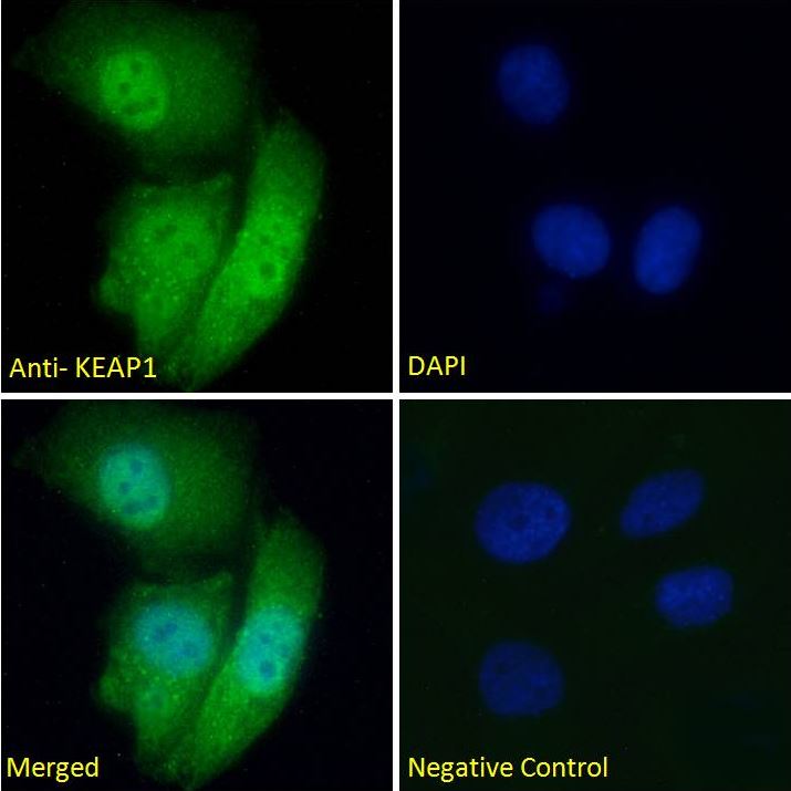
ARG42022 anti-KEAP1 antibody ICC/IF image
Immunofluorescence: HeLa cells fixed with Paraformaldehyde and permeabilized with 0.15% Triton. Cells were stained with ARG42022 anti-KEAP1 antibody (green) at 5 µg/ml dilution for 1 hour. DAPI (blue) for nuclear staining. Negative control: Unimmunized Goat IgG at 5 µg/ml dilution.
-

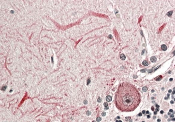
ARG42022 anti-KEAP1 antibody IHC-P image
Immunohistochemistry: Paraffin-embedded Human cerebellum tissue. Antigen Retrieval: Steam tissue section in Citrate buffer (pH 6.0). The tissue section was stained with ARG42022 anti-KEAP1 antibody at 3.8 µg/ml dilution.
-
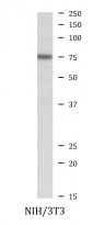
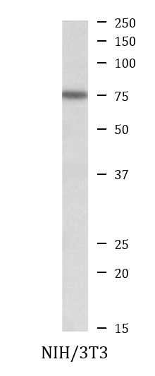
ARG42022 anti-KEAP1 antibody WB image
Western blot: 35 µg of NIH/3T3 cell lysate (in RIPA buffer) stained with ARG42022 anti-KEAP1 antibody at 0.2 µg/ml dilution and incubated at RT for 1 hour.