ARG40406
anti-IRAK1 antibody
anti-IRAK1 antibody for ICC/IF,Western blot and Human
概述
| 产品描述 | Rabbit Polyclonal antibody recognizes IRAK1 |
|---|---|
| 反应物种 | Hu |
| 应用 | ICC/IF, WB |
| 特异性 | At least three isoforms of IRAK1 are known to exist; this antibody will detect all three isoforms. The antibody is predicted to not cross-react with other members of the IRAK protein family. |
| 宿主 | Rabbit |
| 克隆 | Polyclonal |
| 同位型 | IgG |
| 靶点名称 | IRAK1 |
| 抗原物种 | Human |
| 抗原 | A peptide (13 amino acids) within the last 50 amino acids of Human IRAK1. |
| 偶联标记 | Un-conjugated |
| 別名 | Interleukin-1 receptor-associated kinase 1; IRAK; IRAK-1; EC 2.7.11.1; pelle |
应用说明
| 应用建议 |
|
||||||
|---|---|---|---|---|---|---|---|
| 应用说明 | * The dilutions indicate recommended starting dilutions and the optimal dilutions or concentrations should be determined by the scientist. | ||||||
| 阳性对照 | HeLa | ||||||
| 实际分子量 | ~ 80 kDa |
属性
| 形式 | Liquid |
|---|---|
| 纯化 | Affinity purification with immunogen. |
| 缓冲液 | PBS and 0.02% Sodium azide. |
| 抗菌剂 | 0.02% Sodium azide |
| 浓度 | 1 mg/ml |
| 存放说明 | For continuous use, store undiluted antibody at 2-8°C for up to a week. For long-term storage, aliquot and store at -20°C or below. Storage in frost free freezers is not recommended. Avoid repeated freeze/thaw cycles. Suggest spin the vial prior to opening. The antibody solution should be gently mixed before use. |
| 注意事项 | For laboratory research only, not for drug, diagnostic or other use. |
生物信息
| 数据库连接 |
Swiss-port # P51617 Human Interleukin-1 receptor-associated kinase 1 |
|---|---|
| 基因名称 | IRAK1 |
| 全名 | interleukin-1 receptor-associated kinase 1 |
| 背景介绍 | This gene encodes the interleukin-1 receptor-associated kinase 1, one of two putative serine/threonine kinases that become associated with the interleukin-1 receptor (IL1R) upon stimulation. This gene is partially responsible for IL1-induced upregulation of the transcription factor NF-kappa B. Alternatively spliced transcript variants encoding different isoforms have been found for this gene. [provided by RefSeq, Jul 2008] |
| 生物功能 | Serine/threonine-protein kinase that plays a critical role in initiating innate immune response against foreign pathogens. Involved in Toll-like receptor (TLR) and IL-1R signaling pathways. Is rapidly recruited by MYD88 to the receptor-signaling complex upon TLR activation. Association with MYD88 leads to IRAK1 phosphorylation by IRAK4 and subsequent autophosphorylation and kinase activation. Phosphorylates E3 ubiquitin ligases Pellino proteins (PELI1, PELI2 and PELI3) to promote pellino-mediated polyubiquitination of IRAK1. Then, the ubiquitin-binding domain of IKBKG/NEMO binds to polyubiquitinated IRAK1 bringing together the IRAK1-MAP3K7/TAK1-TRAF6 complex and the NEMO-IKKA-IKKB complex. In turn, MAP3K7/TAK1 activates IKKs (CHUK/IKKA and IKBKB/IKKB) leading to NF-kappa-B nuclear translocation and activation. Alternatively, phosphorylates TIRAP to promote its ubiquitination and subsequent degradation. Phosphorylates the interferon regulatory factor 7 (IRF7) to induce its activation and translocation to the nucleus, resulting in transcriptional activation of type I IFN genes, which drive the cell in an antiviral state. When sumoylated, translocates to the nucleus and phosphorylates STAT3. [UniProt] |
| 细胞定位 | Cytoplasm. Nucleus. Lipid droplet. Note=Translocates to the nucleus when sumoylated. RSAD2/viperin recruits it to the lipid droplet (By similarity). [UniProt] |
| 预测分子量 | 77 kDa |
| 翻译后修饰 | Following recruitment on the activated receptor complex, phosphorylated on Thr-209, probably by IRAK4, resulting in a conformational change of the kinase domain, allowing further phosphorylations to take place. Thr-387 phosphorylation in the activation loop is required to achieve full enzymatic activity. Polyubiquitinated by TRAF6 after cell stimulation with IL-1-beta by PELI1, PELI2 and PELI3. Polyubiquitination occurs with polyubiquitin chains linked through 'Lys-63'. Ubiquitination promotes interaction with NEMO/IKBKG. Also sumoylated; leading to nuclear translocation. [UniProt] |
检测图片 (3) Click the Picture to Zoom In
-
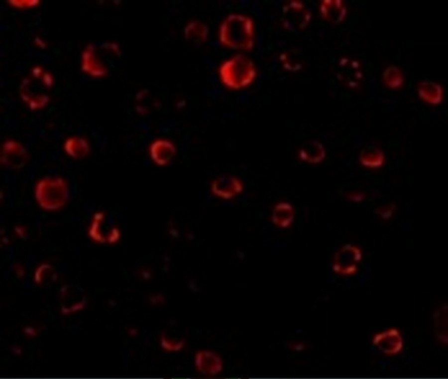
ARG40406 anti-IRAK1 antibody ICC/IF image
Immunofluorescence: HeLa cells stained with ARG40406 anti-IRAK1 antibody at 20 µg/ml dilution.
-
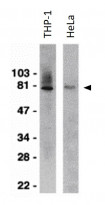
ARG40406 anti-IRAK1 antibody WB image
Western blot: THP-1 and HeLa whole cell lysates stained with ARG40406 anti-IRAK1 antibody at 1 µg/ml dilution.
-
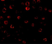
ARG40406 anti-IRAK1 antibody ICC image
Immunocytochemistry: HeLa cells stained with ARG40406 anti-IRAK1 antibody at 10 µg/ml dilution.