ARG10608
anti-IP3 Receptor antibody
anti-IP3 Receptor antibody for Flow cytometry,ICC/IF,IHC-Formalin-fixed paraffin-embedded sections,Western blot and Human,Mouse,Rat
概述
| 产品描述 | Rabbit Polyclonal antibody recognizes IP3 Receptor |
|---|---|
| 反应物种 | Hu, Ms, Rat |
| 应用 | FACS, ICC/IF, IHC-P, WB |
| 宿主 | Rabbit |
| 克隆 | Polyclonal |
| 同位型 | IgG |
| 靶点名称 | IP3 Receptor |
| 抗原物种 | Human |
| 抗原 | Recombinant fragment around aa. 2411-2758 of Human IP3 Receptor. |
| 偶联标记 | Un-conjugated |
| 別名 | IP3R; SCA29; InsP3R1; SCA15; Type 1 InsP3 receptor; SCA16; INSP3R1; PPP1R94; IP3R 1; IP3 receptor isoform 1; ACV; IP3R1; CLA4; Inositol 1,4,5-trisphosphate receptor type 1; Type 1 inositol 1,4,5-trisphosphate receptor |
应用说明
| 应用建议 |
|
||||||||||
|---|---|---|---|---|---|---|---|---|---|---|---|
| 应用说明 | * The dilutions indicate recommended starting dilutions and the optimal dilutions or concentrations should be determined by the scientist. | ||||||||||
| 阳性对照 | SHG-44 (glioma), Rat brain and Mouse brain | ||||||||||
| 实际分子量 | ~ 315 kDa |
属性
| 形式 | Liquid |
|---|---|
| 纯化 | Affinity purification with immunogen. |
| 缓冲液 | PBS, 0.025% Sodium azide and 2.5% BSA. |
| 抗菌剂 | 0.025% Sodium azide |
| 稳定剂 | 2.5% BSA |
| 浓度 | 0.5 mg/ml |
| 存放说明 | For continuous use, store undiluted antibody at 2-8°C for up to a week. For long-term storage, aliquot and store at -20°C or below. Storage in frost free freezers is not recommended. Avoid repeated freeze/thaw cycles. Suggest spin the vial prior to opening. The antibody solution should be gently mixed before use. |
| 注意事项 | For laboratory research only, not for drug, diagnostic or other use. |
生物信息
| 数据库连接 | |
|---|---|
| 基因名称 | ITPR1 |
| 全名 | inositol 1,4,5-trisphosphate receptor, type 1 |
| 背景介绍 | This gene encodes an intracellular receptor for inositol 1,4,5-trisphosphate. Upon stimulation by inositol 1,4,5-trisphosphate, this receptor mediates calcium release from the endoplasmic reticulum. Mutations in this gene cause spinocerebellar ataxia type 15, a disease associated with an heterogeneous group of cerebellar disorders. Multiple transcript variants have been identified for this gene. [provided by RefSeq, Nov 2009] |
| 生物功能 | Intracellular channel that mediates calcium release from the endoplasmic reticulum following stimulation by inositol 1,4,5-trisphosphate. Involved in the regulation of epithelial secretion of electrolytes and fluid through the interaction with AHCYL1 (By similarity). Plays a role in ER stress-induced apoptosis. Cytoplasmic calcium released from the ER triggers apoptosis by the activation of CaM kinase II, eventually leading to the activation of downstream apoptosis pathways (By similarity). [UniProt] |
| 预测分子量 | 314 kDa |
| 翻译后修饰 | Phosphorylated on tyrosine residues. Ubiquitination at multiple lysines targets ITPR1 for proteasomal degradation. Approximately 40% of the ITPR1-associated ubiquitin is monoubiquitin, and polyubiquitins are both 'Lys-48'- and 'Lys-63'-linked (By similarity). Phosphorylated by cAMP kinase (PKA). Phosphorylation prevents the ligand-induced opening of the calcium channels. Phosphorylation by PKA increases the interaction with inositol 1,4,5-trisphosphate and decreases the interaction with AHCYL1. |
检测图片 (8) Click the Picture to Zoom In
-

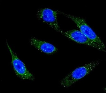
ARG10608 anti-IP3 Receptor antibody ICC/IF image
Immunofluorescence: U-2 OS cells stained with ARG10608 anti-IP3 Receptor antibody (green). DAPI (blue) for nuclear staining.
-
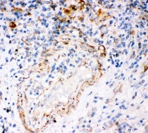
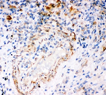
ARG10608 anti-IP3 Receptor antibody IHC-P image
Immunohistochemistry: Human lung cancer tissue stained with ARG10608 anti-IP3 Receptor antibody.
-
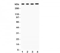
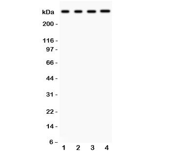
ARG10608 anti-IP3 Receptor antibody WB image
Western blot: 1) Rat brain, 2) Rat liver, 3) HeLa, and 4) HepG2 lysates stained with ARG10608 anti-IP3 Receptor antibody.
-
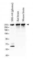
ARG10608 anti-IP3 Receptor antibody WB image
Western blot: SHG-44 (glioma), Rat brain and Mouse brain lysates stained with ARG10608 anti-IP3 Receptor antibody at 1 µg/ml dilution.
-
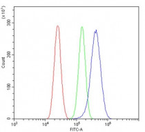
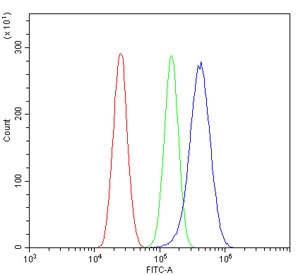
ARG10608 anti-IP3 Receptor antibody FACS image
Flow Cytometry: U-87 MG cells were blocked with goat sera and stained with ARG10608 anti-IP3 Receptor antibody at 1 µg/10^6 cells (blue); Cells alone (red); Isotype control (green).
-

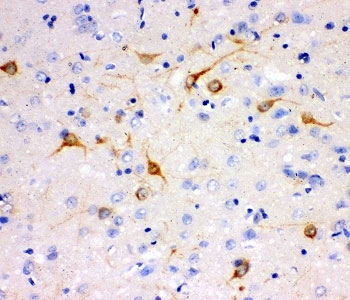
ARG10608 anti-IP3 Receptor antibody IHC-P image
Immunohistochemistry: Mouse brain tissue stained with ARG10608 anti-IP3 Receptor antibody.
-
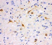
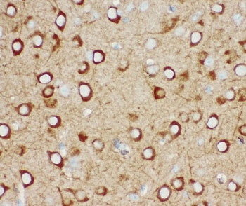
ARG10608 anti-IP3 Receptor antibody IHC-P image
Immunohistochemistry: Rat brain tissue stained with ARG10608 anti-IP3 Receptor antibody.
-
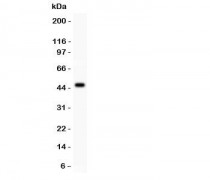
ARG10608 anti-IP3 Receptor antibody WB image
Western blot: 0.5 ng of recombinant human protein stained with ARG10608 anti-IP3 Receptor antibody.