ARG40706
anti-IKZF1 / Ikaros antibody
anti-IKZF1 / Ikaros antibody for Flow cytometry,ICC/IF,IHC-Formalin-fixed paraffin-embedded sections,Western blot and Human,Mouse,Rat
概述
| 产品描述 | Rabbit Polyclonal antibody recognizes IKZF1 / Ikaros |
|---|---|
| 反应物种 | Hu, Ms, Rat |
| 应用 | FACS, ICC/IF, IHC-P, WB |
| 宿主 | Rabbit |
| 克隆 | Polyclonal |
| 同位型 | IgG |
| 靶点名称 | IKZF1 / Ikaros |
| 抗原物种 | Human |
| 抗原 | Synthetic peptide corresponding to aa. 428-459 of Human Ikaros. (LKEEHRAYDLLRAASENSQDALRVVSTSGEQM) |
| 偶联标记 | Un-conjugated |
| 別名 | IK1; Hs.54452; LYF1; PPP1R92; LyF-1; Lymphoid transcription factor LyF-1; IKAROS; Ikaros family zinc finger protein 1; DNA-binding protein Ikaros; PRO0758; ZNFN1A1 |
应用说明
| 应用建议 |
|
||||||||||
|---|---|---|---|---|---|---|---|---|---|---|---|
| 应用说明 | * The dilutions indicate recommended starting dilutions and the optimal dilutions or concentrations should be determined by the scientist. |
属性
| 形式 | Liquid |
|---|---|
| 纯化 | Affinity purification with immunogen. |
| 缓冲液 | 0.2% Na2HPO4, 0.9% NaCl, 0.05% Sodium azide and 5% BSA. |
| 抗菌剂 | 0.05% Sodium azide |
| 稳定剂 | 5% BSA |
| 浓度 | 0.5 mg/ml |
| 存放说明 | For continuous use, store undiluted antibody at 2-8°C for up to a week. For long-term storage, aliquot and store at -20°C or below. Storage in frost free freezers is not recommended. Avoid repeated freeze/thaw cycles. Suggest spin the vial prior to opening. The antibody solution should be gently mixed before use. |
| 注意事项 | For laboratory research only, not for drug, diagnostic or other use. |
生物信息
| 数据库连接 | |
|---|---|
| 基因名称 | IKZF1 |
| 全名 | IKAROS family zinc finger 1 (Ikaros) |
| 背景介绍 | This gene encodes a transcription factor that belongs to the family of zinc-finger DNA-binding proteins associated with chromatin remodeling. The expression of this protein is restricted to the fetal and adult hemo-lymphopoietic system, and it functions as a regulator of lymphocyte differentiation. Several alternatively spliced transcript variants encoding different isoforms have been described for this gene. Most isoforms share a common C-terminal domain, which contains two zinc finger motifs that are required for hetero- or homo-dimerization, and for interactions with other proteins. The isoforms, however, differ in the number of N-terminal zinc finger motifs that bind DNA and in nuclear localization signal presence, resulting in members with and without DNA-binding properties. Only a few isoforms contain the requisite three or more N-terminal zinc motifs that confer high affinity binding to a specific core DNA sequence element in the promoters of target genes. The non-DNA-binding isoforms are largely found in the cytoplasm, and are thought to function as dominant-negative factors. Overexpression of some dominant-negative isoforms have been associated with B-cell malignancies, such as acute lymphoblastic leukemia (ALL). [provided by RefSeq, May 2014] |
| 生物功能 | Transcription regulator of hematopoietic cell differentiation. Binds gamma-satellite DNA. Plays a role in the development of lymphocytes, B- and T-cells. Binds and activates the enhancer (delta-A element) of the CD3-delta gene. Repressor of the TDT (fikzfterminal deoxynucleotidyltransferase) gene during thymocyte differentiation. Regulates transcription through association with both HDAC-dependent and HDAC-independent complexes. Targets the 2 chromatin-remodeling complexes, NuRD and BAF (SWI/SNF), in a single complex (PYR complex), to the beta-globin locus in adult erythrocytes. Increases normal apoptosis in adult erythroid cells. Confers early temporal competence to retinal progenitor cells (RPCs) (By similarity). Function is isoform-specific and is modulated by dominant-negative inactive isoforms. [UniProt] |
| 细胞定位 | Nucleus. Isoform Ik2: Nucleus. Isoform Ik6: Cytoplasm. [UniProt] |
| 预测分子量 | 58 kDa |
| 翻译后修饰 | Phosphorylation controls cell-cycle progression from late G(1) stage to S stage. Hyperphosphorylated during G2/M phase. Dephosphorylated state during late G(1) phase. Phosphorylation on Thr-140 is required for DNA and pericentromeric location during mitosis. CK2 is the main kinase, in vitro. GSK3 and CDK may also contribute to phosphorylation of the C-terminal serine and threonine residues. Phosphorylation on these C-terminal residues reduces the DNA-binding ability. Phosphorylation/dephosphorylation events on Ser-13 and Ser-295 regulate TDT expression during thymocyte differentiation. Dephosphorylation by protein phosphatase 1 regulates stability and pericentromeric heterochromatin location. Phosphorylated in both lymphoid and non-lymphoid tissues (By similarity). Phosphorylation at Ser-361 and Ser-364 downstream of SYK induces nuclear translocation. Sumoylated. Simulataneous sumoylation on the 2 sites results in a loss of both HDAC-dependent and HDAC-independent repression. Has no effect on pericentromeric heterochromatin location. Desumoylated by SENP1 (By similarity). Polyubiquitinated. [UniProt] |
检测图片 (6) Click the Picture to Zoom In
-
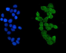
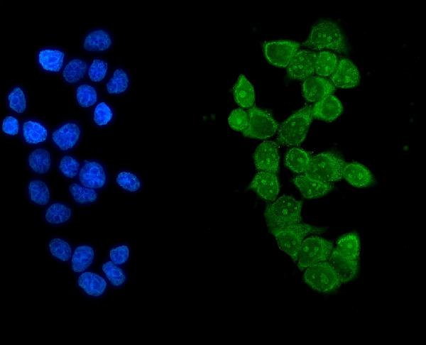
ARG40706 anti-IKZF1 / Ikaros antibody ICC/IF image
Immunofluorescence: MCF-7 cells were blocked with 10% goat serum and then stained with ARG40706 anti-IKZF1 / Ikaros antibody (green) at 5 µg/ml dilution, overnight at 4°C. DAPI (blue) for nuclear staining.
-
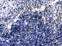
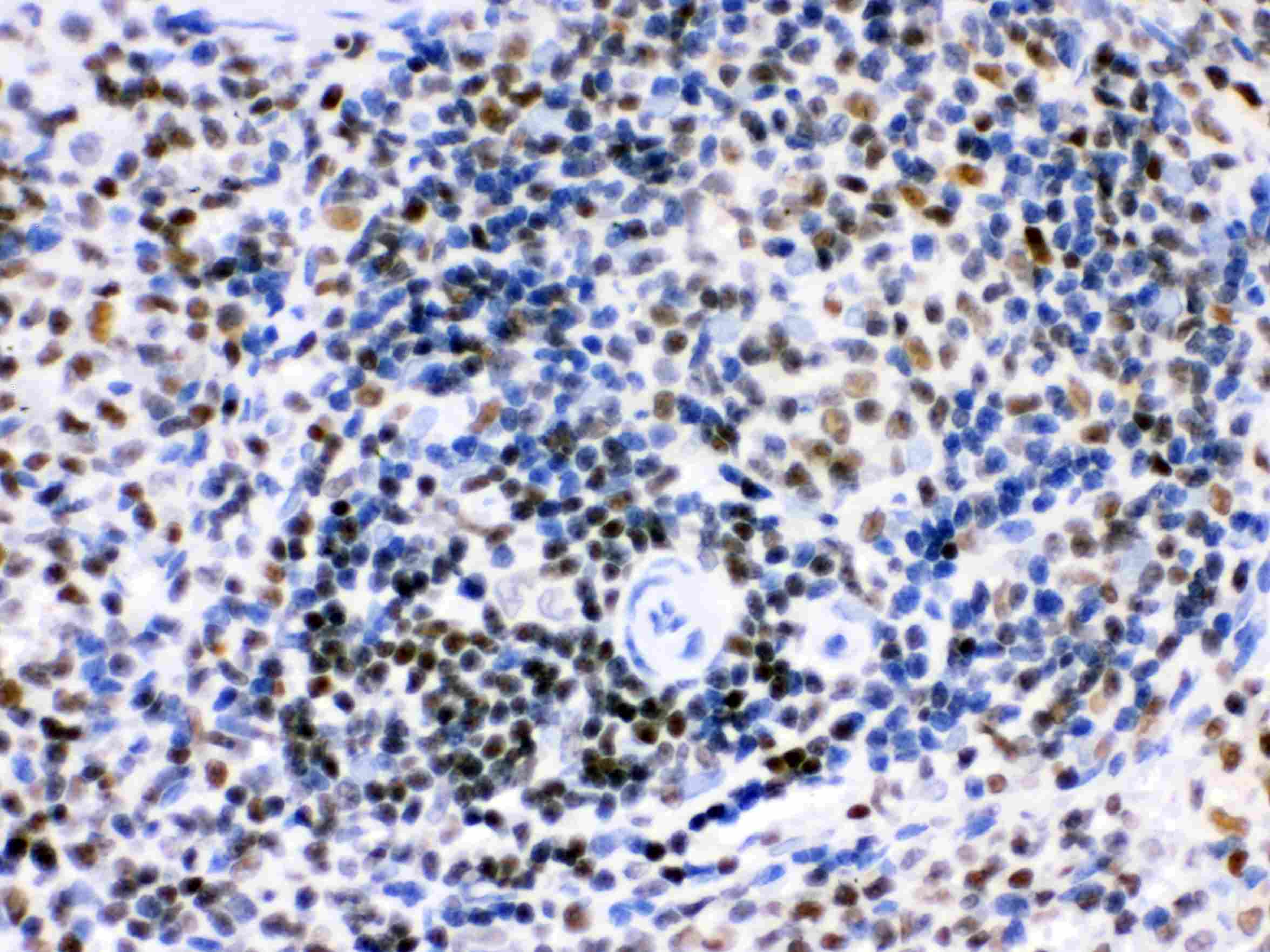
ARG40706 anti-IKZF1 / Ikaros antibody IHC-P image
Immunohistochemistry: Paraffin-embedded Mouse spleen tissue stained with ARG40706 anti-IKZF1 / Ikaros antibody.
-
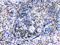
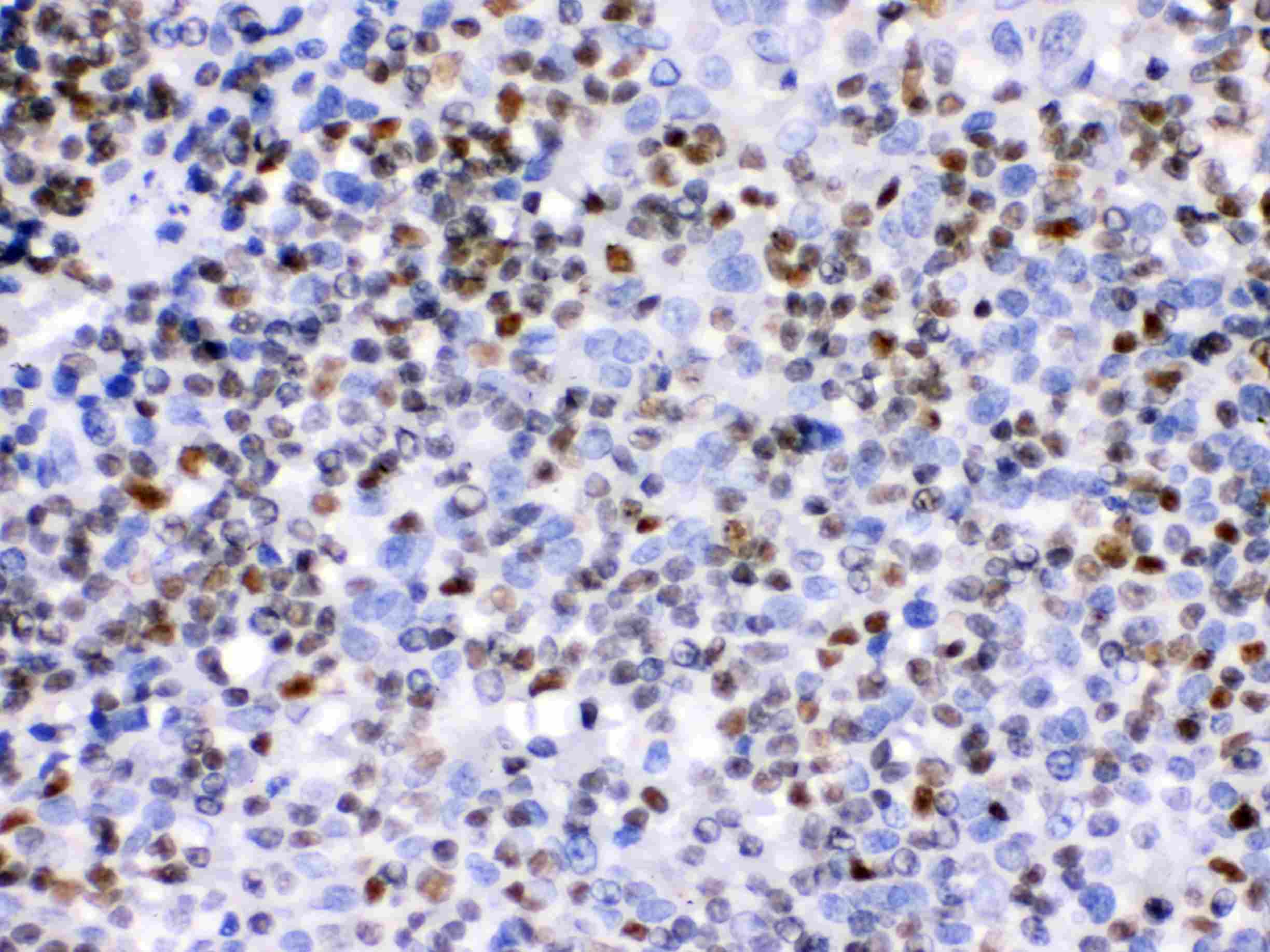
ARG40706 anti-IKZF1 / Ikaros antibody IHC-P image
Immunohistochemistry: Paraffin-embedded Rat spleen tissue stained with ARG40706 anti-IKZF1 / Ikaros antibody.
-

ARG40706 anti-IKZF1 / Ikaros antibody IHC-P image
Immunohistochemistry: Paraffin-embedded Human tonsil tissue stained with ARG40706 anti-IKZF1 / Ikaros antibody.
-
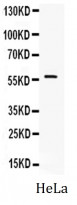
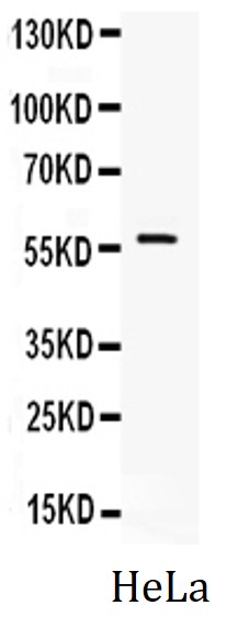
ARG40706 anti-IKZF1 / Ikaros antibody WB image
Western blot: 40 µg of HeLa whole cell lysate stained with ARG40706 anti-IKZF1 / Ikaros antibody at 0.5 µg/ml dilution.
-
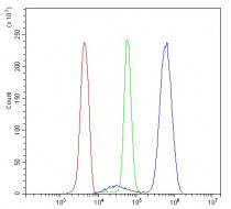
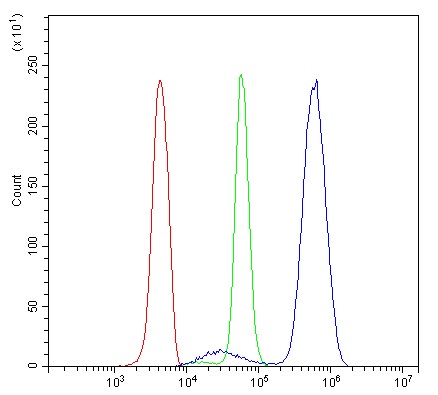
ARG40706 anti-IKZF1 / Ikaros antibody FACS image
Flow Cytometry: U937 cells were blocked with 10% normal goat serum and then stained with ARG40706 anti-IKZF1 / Ikaros antibody (blue) at 1 µg/10^6 cells for 30 min at 20°C, followed by incubation with DyLight®488 labelled secondary antibody. Isotype control antibody (green) was rabbit IgG (1 µg/10^6 cells) used under the same conditions. Unlabelled sample (red) was also used as a control.