ARG42175
anti-HLA DQA2 antibody
anti-HLA DQA2 antibody for IHC-Formalin-fixed paraffin-embedded sections,Western blot and Human
概述
| 产品描述 | Goat Polyclonal antibody recognizes HLA DQA2 |
|---|---|
| 反应物种 | Hu |
| 应用 | IHC-P, WB |
| 宿主 | Goat |
| 克隆 | Polyclonal |
| 同位型 | IgG |
| 靶点名称 | HLA DQA2 |
| 抗原物种 | Human |
| 抗原 | Synthetic peptide around the internal region of Human HLA DQA2. (C-QSHGPSGQYTH) (NP_064440.1) |
| 偶联标记 | Un-conjugated |
| 別名 | MHC class II DQA2; DX alpha chain; HLA class II histocompatibility antigen, DQ; HLA class II histocompatibility antigen, DQ alpha 2 chain; HLA-DXA; 6; HLA-DQA1; DX-ALPHA |
应用说明
| 应用建议 |
|
||||||
|---|---|---|---|---|---|---|---|
| 应用说明 | WB: Recommend incubate at RT for 1h. IHC-P: Antigen Retrieval: Steam tissue section in Citrate buffer (pH 6.0). * The dilutions indicate recommended starting dilutions and the optimal dilutions or concentrations should be determined by the scientist. |
||||||
| 阳性对照 | Human bone marrow | ||||||
| 实际分子量 | ~ 26 kDa |
属性
| 形式 | Liquid |
|---|---|
| 纯化 | Affinity purified |
| 缓冲液 | Tris saline (pH 7.3), 0.02% Sodium azide and 0.5% BSA. |
| 抗菌剂 | 0.02% Sodium azide |
| 稳定剂 | 0.5% BSA |
| 浓度 | 0.5 mg/ml |
| 存放说明 | For continuous use, store undiluted antibody at 2-8°C for up to a week. For long-term storage, aliquot and store at -20°C or below. Storage in frost free freezers is not recommended. Avoid repeated freeze/thaw cycles. Suggest spin the vial prior to opening. The antibody solution should be gently mixed before use. |
| 注意事项 | For laboratory research only, not for drug, diagnostic or other use. |
生物信息
| 数据库连接 |
Swiss-port # P01906 Human HLA class II histocompatibility antigen, DQ alpha 2 chain |
|---|---|
| 基因名称 | HLA-DQA2 |
| 全名 | major histocompatibility complex, class II, DQ alpha 2 |
| 背景介绍 | This gene belongs to the HLA class II alpha chain family. The encoded protein forms a heterodimer with a class II beta chain. It is located in intracellular vesicles and plays a central role in the peptide loading of MHC class II molecules by helping to release the CLIP molecule from the peptide binding site. Class II molecules are expressed in antigen presenting cells (B lymphocytes, dendritic cells, macrophages) and are used to present antigenic peptides on the cell surface to be recognized by CD4 T-cells. [provided by RefSeq, Jun 2010] |
| 生物功能 | Binds peptides derived from antigens that access the endocytic route of antigen presenting cells (APC) and presents them on the cell surface for recognition by the CD4 T-cells. The peptide binding cleft accommodates peptides of 10-30 residues. The peptides presented by MHC class II molecules are generated mostly by degradation of proteins that access the endocytic route, where they are processed by lysosomal proteases and other hydrolases. Exogenous antigens that have been endocytosed by the APC are thus readily available for presentation via MHC II molecules, and for this reason this antigen presentation pathway is usually referred to as exogenous. As membrane proteins on their way to degradation in lysosomes as part of their normal turn-over are also contained in the endosomal/lysosomal compartments, exogenous antigens must compete with those derived from endogenous components. Autophagy is also a source of endogenous peptides, autophagosomes constitutively fuse with MHC class II loading compartments. In addition to APCs, other cells of the gastrointestinal tract, such as epithelial cells, express MHC class II molecules and CD74 and act as APCs, which is an unusual trait of the GI tract. To produce a MHC class II molecule that presents an antigen, three MHC class II molecules (heterodimers of an alpha and a beta chain) associate with a CD74 trimer in the ER to form a heterononamer. Soon after the entry of this complex into the endosomal/lysosomal system where antigen processing occurs, CD74 undergoes a sequential degradation by various proteases, including CTSS and CTSL, leaving a small fragment termed CLIP (class-II-associated invariant chain peptide). The removal of CLIP is facilitated by HLA-DM via direct binding to the alpha-beta-CLIP complex so that CLIP is released. HLA-DM stabilizes MHC class II molecules until primary high affinity antigenic peptides are bound. The MHC II molecule bound to a peptide is then transported to the cell membrane surface. In B-cells, the interaction between HLA-DM and MHC class II molecules is regulated by HLA-DO. Primary dendritic cells (DCs) also to express HLA-DO. Lysosomal microenvironment has been implicated in the regulation of antigen loading into MHC II molecules, increased acidification produces increased proteolysis and efficient peptide loading. [UniProt] |
| 细胞定位 | Cell membrane; Single-pass type I membrane protein. Endoplasmic reticulum membrane; Single-pass type I membrane protein. Golgi apparatus, trans-Golgi network membrane; Single-pass type I membrane protein. Endosome membrane; Single-pass type I membrane protein. Lysosome membrane; Single-pass type I membrane protein. Note=The MHC class II complex transits through a number of intracellular compartments in the endocytic pathway until it reaches the cell membrane for antigen presentation. [UniProt] |
| 预测分子量 | 28 kDa |
检测图片 (2) Click the Picture to Zoom In
-
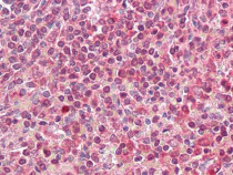
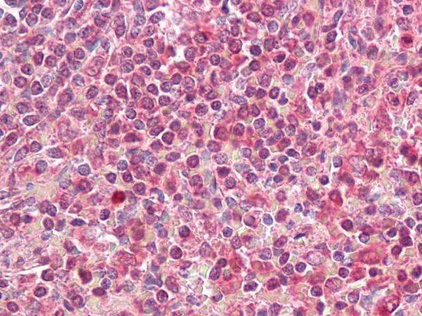
ARG42175 anti-HLA DQA2 antibody IHC-P image
Immunohistochemistry: Paraffin-embedded Human spleen tissue. Antigen Retrieval: Steam tissue section in Citrate buffer (pH 6.0). The tissue section was stained with ARG42175 anti-HLA DQA2 antibody at 3.75 µg/ml dilution followed by AP-staining.
-
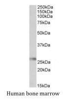
ARG42175 anti-HLA DQA2 antibody WB image
Western blot: 35 µg of Human bone marrow lysate (in RIPA buffer) stained with ARG42175 anti-HLA DQA2 antibody at 0.3 µg/ml dilution and incubated at RT for 1 hour.