ARG58003
anti-HINT1 antibody [1500CT836.13.93]
anti-HINT1 antibody [1500CT836.13.93] for Flow cytometry,IHC-Formalin-fixed paraffin-embedded sections,Western blot and Human,Mouse,Rat
概述
| 产品描述 | Mouse Monoclonal antibody recognizes HINT1 |
|---|---|
| 反应物种 | Hu, Ms, Rat |
| 应用 | FACS, IHC-P, WB |
| 宿主 | Mouse |
| 克隆 | Monoclonal |
| 克隆号 | 1500CT836.13.93 |
| 同位型 | IgG1, kappa |
| 靶点名称 | HINT1 |
| 抗原物种 | Human |
| 抗原 | Recombinant protein of Human HINT1. |
| 偶联标记 | Un-conjugated |
| 別名 | Adenosine 5'-monophosphoramidase; Protein kinase C inhibitor 1; PKCI-1; Protein kinase C-interacting protein 1; HINT; NMAN; EC 3.-.-.-; Histidine triad nucleotide-binding protein 1; PRKCNH1 |
应用说明
| 应用建议 |
|
||||||||
|---|---|---|---|---|---|---|---|---|---|
| 应用说明 | IHC-P: Antigen Retrieval: Heat mediation was performed in Citrate buffer (pH 6.0). * The dilutions indicate recommended starting dilutions and the optimal dilutions or concentrations should be determined by the scientist. |
||||||||
| 阳性对照 | Jurkat | ||||||||
| 实际分子量 | ~ 15 kDa |
属性
| 形式 | Liquid |
|---|---|
| 纯化 | Purification with Protein G. |
| 缓冲液 | PBS and 0.09% (W/V) Sodium azide. |
| 抗菌剂 | 0.09% (W/V) Sodium azide. |
| 存放说明 | For continuous use, store undiluted antibody at 2-8°C for up to a week. For long-term storage, aliquot and store at -20°C or below. Storage in frost free freezers is not recommended. Avoid repeated freeze/thaw cycles. Suggest spin the vial prior to opening. The antibody solution should be gently mixed before use. |
| 注意事项 | For laboratory research only, not for drug, diagnostic or other use. |
生物信息
| 数据库连接 | |
|---|---|
| 基因名称 | HINT1 |
| 全名 | histidine triad nucleotide binding protein 1 |
| 背景介绍 | The protein encoded by this gene can hydrolyze substrates such as AMP-morpholidate, AMP-N-alanine methyl ester, AMP-alpha-acetyl lysine methyl ester, and AMP-NH2. The encoded protein interacts with these substrates via a histidine triad motif, which is part of the loop that binds to the substrate. This gene has been found to be a tumor suppressing gene. Several transcript variants, but only one of them protein-coding, have been found for this gene. [provided by RefSeq, Dec 2012] |
| 生物功能 | Hydrolyzes purine nucleotide phosphoramidates with a single phosphate group, including adenosine 5'monophosphoramidate (AMP-NH2), adenosine 5'monophosphomorpholidate (AMP-morpholidate) and guanosine 5'monophosphomorpholidate (GMP-morpholidate). Hydrolyzes lysyl-AMP (AMP-N-epsilon-(N-alpha-acetyl lysine methyl ester)) generated by lysine tRNA ligase, as well as Met-AMP, His-AMP and Asp-AMP, lysyl-GMP (GMP-N-epsilon-(N-alpha-acetyl lysine methyl ester)) and AMP-N-alanine methyl ester. Can also convert adenosine 5'-O-phosphorothioate and guanosine 5'-O-phosphorothioate to the corresponding nucleoside 5'-O-phosphates with concomitant release of hydrogen sulfide. In addition, functions as scaffolding protein that modulates transcriptional activation by the LEF1/TCF1-CTNNB1 complex and by the complex formed with MITF and CTNNB1. Modulates p53/TP53 levels and p53/TP53-mediated apoptosis. Modulates proteasomal degradation of target proteins by the SCF (SKP2-CUL1-F-box protein) E3 ubiquitin-protein ligase complex. [UniProt] |
| 预测分子量 | 14 kDa |
检测图片 (3) Click the Picture to Zoom In
-
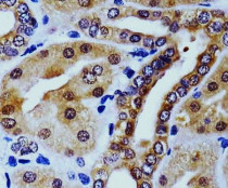
ARG58003 anti-HINT1 antibody IHC-P image
Immunohistochemistry: Paraffin-embedded Human kidney tissue was fixed with formaldehyde and blocked with 3% BSA for 0.5 hour at RT. Samples were stained with ARG58003 anti-HINT1 antibody at 1:25 dilution for 60 min at 37°C. Antigen Retrieval: Heat mediation was performed in Citrate buffer (pH 6.0).
-
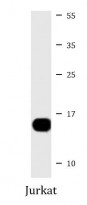
ARG58003 anti-HINT1 antibody WB image
Western blot: 20 µg of Jurkat cell lysate stained with ARG58003 anti-HINT1 antibody at 1:4000 dilution.
-
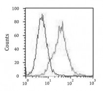
ARG58003 anti-HINT1 antibody FACS image
Flow Cytometry: Jurkat cells were fixed with 2% paraformaldehyde (10 min) and then permeabilized with 90% methanol for 10 min. Cells were then incubated in 2% BSA to block non-specific protein-protein interactions and stained with ARG58003 anti-HINT1 antibody (right histogram) at 1:25 dilution for 60 min at 37°C, followed by DyLight® 488 labelled secondary antibody. Isotype control antibody (left histogram) was Mouse IgG1 (1 µg/10^6 cells) used under the same conditions.


