ARG66319
anti-HDAC1 antibody [SQab1867]
anti-HDAC1 antibody [SQab1867] for Flow cytometry,ICC/IF,IHC-Formalin-fixed paraffin-embedded sections,Western blot and Human,Bovine,Monkey
概述
| 产品描述 | Recombinant Rabbit Monoclonal antibody [SQab1867] recognizes HDAC1 |
|---|---|
| 反应物种 | Hu, Bov, Mk |
| 应用 | FACS, ICC/IF, IHC-P, WB |
| 宿主 | Rabbit |
| 克隆 | Monoclonal |
| 克隆号 | SQab1867 |
| 同位型 | IgG |
| 靶点名称 | HDAC1 |
| 抗原物种 | Human |
| 抗原 | Synthetic peptide around the C-terminus of HDAC1. |
| 偶联标记 | Un-conjugated |
| 別名 | EC 3.5.1.98; HD1; RPD3L1; Histone deacetylase 1; GON-10; RPD3 |
应用说明
| 应用建议 |
|
||||||||||
|---|---|---|---|---|---|---|---|---|---|---|---|
| 应用说明 | IHC-P: Antigen Retrieval: Heat mediated was performed using Tris/EDTA buffer pH 9.0. * The dilutions indicate recommended starting dilutions and the optimal dilutions or concentrations should be determined by the scientist. |
属性
| 形式 | Liquid |
|---|---|
| 纯化 | Purification with Protein A. |
| 缓冲液 | PBS, 0.01% Sodium azide, 40% Glycerol and 0.05% BSA. |
| 抗菌剂 | 0.01% Sodium azide |
| 稳定剂 | 40% Glycerol and 0.05% BSA |
| 存放说明 | For continuous use, store undiluted antibody at 2-8°C for up to a week. For long-term storage, aliquot and store at -20°C. Storage in frost free freezers is not recommended. Avoid repeated freeze/thaw cycles. Suggest spin the vial prior to opening. The antibody solution should be gently mixed before use. |
| 注意事项 | For laboratory research only, not for drug, diagnostic or other use. |
生物信息
| 数据库连接 | |
|---|---|
| 基因名称 | HDAC1 |
| 全名 | histone deacetylase 1 |
| 背景介绍 | Histone acetylation and deacetylation, catalyzed by multisubunit complexes, play a key role in the regulation of eukaryotic gene expression. The protein encoded by this gene belongs to the histone deacetylase/acuc/apha family and is a component of the histone deacetylase complex. It also interacts with retinoblastoma tumor-suppressor protein and this complex is a key element in the control of cell proliferation and differentiation. Together with metastasis-associated protein-2, it deacetylates p53 and modulates its effect on cell growth and apoptosis. [provided by RefSeq, Jul 2008] |
| 生物功能 | Responsible for the deacetylation of lysine residues on the N-terminal part of the core histones (H2A, H2B, H3 and H4). Histone deacetylation gives a tag for epigenetic repression and plays an important role in transcriptional regulation, cell cycle progression and developmental events. Histone deacetylases act via the formation of large multiprotein complexes. Deacetylates SP proteins, SP1 and SP3, and regulates their function. Component of the BRG1-RB1-HDAC1 complex, which negatively regulates the CREST-mediated transcription in resting neurons. Upon calcium stimulation, HDAC1 is released from the complex and CREBBP is recruited, which facilitates transcriptional activation. Deacetylates TSHZ3 and regulates its transcriptional repressor activity. Deacetylates 'Lys-310' in RELA and thereby inhibits the transcriptional activity of NF-kappa-B. Deacetylates NR1D2 and abrogates the effect of KAT5-mediated relieving of NR1D2 transcription repression activity. Component of a RCOR/GFI/KDM1A/HDAC complex that suppresses, via histone deacetylase (HDAC) recruitment, a number of genes implicated in multilineage blood cell development. Involved in CIART-mediated transcriptional repression of the circadian transcriptional activator: CLOCK-ARNTL/BMAL1 heterodimer. Required for the transcriptional repression of circadian target genes, such as PER1, mediated by the large PER complex or CRY1 through histone deacetylation. [UniProt] |
| 产品亮点 | Related products: HDAC1 antibodies; Anti-Rabbit IgG secondary antibodies; Related news: Cancer Pathology Markers (SQ clones) |
| 预测分子量 | 55 kDa |
| 翻译后修饰 | Sumoylated on Lys-444 and Lys-476; which promotes enzymatic activity. Desumoylated by SENP1. Phosphorylation on Ser-421 and Ser-423 promotes enzymatic activity and interactions with NuRD and SIN3 complexes. Phosphorylated by CDK5. Ubiquitinated by CHFR, leading to its degradation by the proteasome. Ubiquitinated by KCTD11, leading to proteasomal degradation. [UniProt] |
检测图片 (5) Click the Picture to Zoom In
-

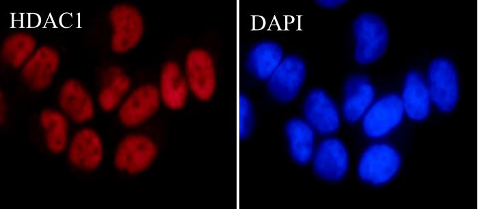
ARG66319 anti-HDAC1 antibody [SQab1867] ICC/IF image
Immunofluorescence: HeLa cells were fixed with 4% paraformaldehyde for 30 min at RT, permeabilized with 0.1% Triton X-100 for 10 min at RT then blocked with 10% goat serum for 30 min at RT. Cells were stained with ARG66319 anti-HDAC1 antibody [SQab1867] (red) at 1:500 and 4°C. DAPI (blue) was used as the nuclear counter stain.
-
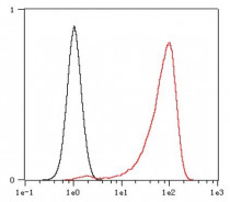
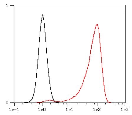
ARG66319 anti-HDAC1 antibody [SQab1867] FACS image
Flow Cytometry: HeLa cells were fixed with 4% paraformaldehyde (10 min) and then permeabilized with 0.1% TritonX-100 for 15 min. The cells were stained with ARG66319 anti-HDAC1 antibody [SQab1867] (red) at 1:200 dilution in 1x PBS/1% BSA for 30 min at RT, followed by Alexa Fluor® 488 labelled secondary antibody. Unlabelled sample (black) was used as a control.
-
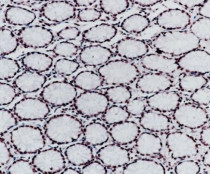
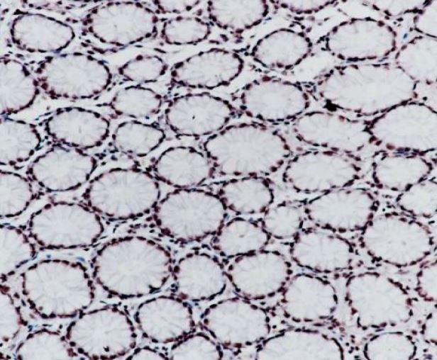
ARG66319 anti-HDAC1 antibody [SQab1867] IHC-P image
Immunohistochemistry: Formalin-fixed and paraffin-embedded Human colon tissue stained with ARG66319 anti-HDAC1 antibody [SQab1867] at 1:200 dilution. Antigen Retrieval: Heat mediated was performed using Tris/EDTA buffer pH 9.0.
-
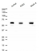
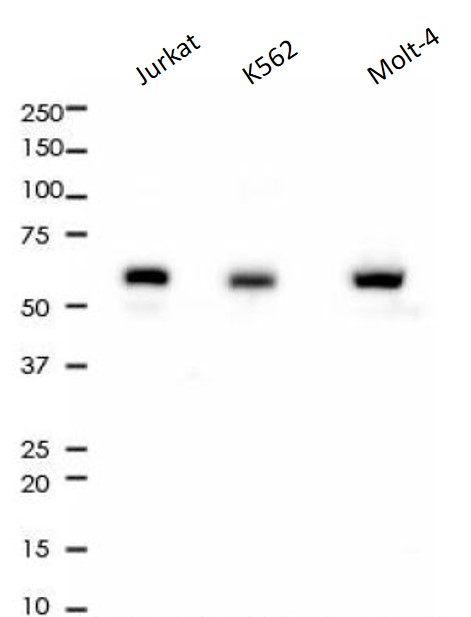
ARG66319 anti-HDAC1 antibody [SQab1867] WB image
Western blot: 10 µg of Jurkat, K562 and Molt-4 cell lysates stained with ARG66319 anti-HDAC1 antibody [SQab1867] at 1:1000 dilution.
-

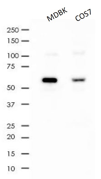
ARG66319 anti-HDAC1 antibody [SQab1867] WB image
Western blot: 10 µg of MDBK and COS7 cell lysates stained with ARG66319 anti-HDAC1 antibody [SQab1867] at 1:1000 dilution.