ARG42839
anti-Glypican 5 antibody
anti-Glypican 5 antibody for Flow cytometry,ICC/IF,Western blot and Human
概述
| 产品描述 | Rabbit Polyclonal antibody recognizes Glypican 5 |
|---|---|
| 反应物种 | Hu |
| 应用 | FACS, ICC/IF, WB |
| 宿主 | Rabbit |
| 克隆 | Polyclonal |
| 同位型 | IgG |
| 靶点名称 | Glypican 5 |
| 抗原物种 | Human |
| 抗原 | Recombinant protein corresponding to Q28-D538 of Human Glypican 5. |
| 偶联标记 | Un-conjugated |
| 別名 | Glypican-5 |
应用说明
| 应用建议 |
|
||||||||
|---|---|---|---|---|---|---|---|---|---|
| 应用说明 | * The dilutions indicate recommended starting dilutions and the optimal dilutions or concentrations should be determined by the scientist. | ||||||||
| 实际分子量 | ~ 64 kDa |
属性
| 形式 | Liquid |
|---|---|
| 纯化 | Affinity purification with immunogen. |
| 缓冲液 | 0.2% Na2HPO4, 0.9% NaCl, 0.01% Sodium azide and 4% Trehalose. |
| 抗菌剂 | 0.01% Sodium azide |
| 稳定剂 | 4% Trehalose |
| 浓度 | 0.5 mg/ml |
| 存放说明 | For continuous use, store undiluted antibody at 2-8°C for up to a week. For long-term storage, aliquot and store at -20°C or below. Storage in frost free freezers is not recommended. Avoid repeated freeze/thaw cycles. Suggest spin the vial prior to opening. The antibody solution should be gently mixed before use. |
| 注意事项 | For laboratory research only, not for drug, diagnostic or other use. |
生物信息
| 数据库连接 | |
|---|---|
| 基因名称 | GPC5 |
| 全名 | glypican 5 |
| 背景介绍 | Cell surface heparan sulfate proteoglycans are composed of a membrane-associated protein core substituted with a variable number of heparan sulfate chains. Members of the glypican-related integral membrane proteoglycan family (GRIPS) contain a core protein anchored to the cytoplasmic membrane via a glycosyl phosphatidylinositol linkage. These proteins may play a role in the control of cell division and growth regulation. [provided by RefSeq, Jul 2008] |
| 生物功能 | Cell surface proteoglycan that bears heparan sulfate. [UniProt] |
| 细胞定位 | Cell membrane; Lipid-anchor, GPI-anchor; Extracellular side. Secreted glypican-5: Secreted, extracellular space. [UniProt] |
| 预测分子量 | 64 kDa |
检测图片 (4) Click the Picture to Zoom In
-
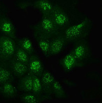
ARG42839 anti-Glypican 5 antibody ICC/IF image
Immunofluorescence: A431 cells were blocked with 10% goat serum and then stained with ARG42839 anti-Glypican 5 antibody (green) at 4 µg/ml dilution, overnight at 4°C.
-
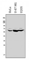
ARG42839 anti-Glypican 5 antibody WB image
Western blot: 50 µg of sample under reducing conditions. HeLa, U-87 MG and U2OS whole cell lysates stained with ARG42839 anti-Glypican 5 antibody at 0.5 µg/ml dilution, overnight at 4°C.
-
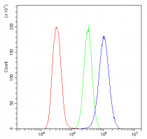
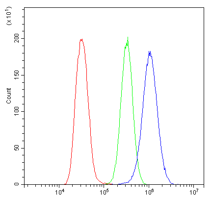
ARG42839 anti-Glypican 5 antibody FACS image
Flow Cytometry: U2OS cells were blocked with 10% normal goat serum and then stained with ARG42839 anti-Glypican 5 antibody (blue) at 1 µg/10^6 cells for 30 min at 20°C, followed by incubation with DyLight®488 labelled secondary antibody. Isotype control antibody (green) was Rabbit IgG (1 µg/10^6 cells) used under the same conditions. Unlabelled sample (red) was also used as a control.
-
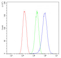
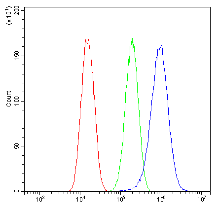
ARG42839 anti-Glypican 5 antibody FACS image
Flow Cytometry: U-87 MG cells were blocked with 10% normal goat serum and then stained with ARG42839 anti-Glypican 5 antibody (blue) at 1 µg/10^6 cells for 30 min at 20°C, followed by incubation with DyLight®488 labelled secondary antibody. Isotype control antibody (green) was Rabbit IgG (1 µg/10^6 cells) used under the same conditions. Unlabelled sample (red) was also used as a control.