ARG62993
anti-GFP antibody
anti-GFP antibody for ICC/IF,Immunoprecipitation,Western blot and Other
Controls and Markers antibody; Tag Internal Control antibody; Fluorescent-Tags antibody
概述
| 产品描述 | Rabbit Polyclonal antibody recognizes GFP |
|---|---|
| 反应物种 | Other |
| 应用 | ICC/IF, IP, WB |
| 特异性 | The polyclonal antibody recognizes GFP, EGFP, EYFP fusion proteins in all species. |
| 宿主 | Rabbit |
| 克隆 | Polyclonal |
| 同位型 | IgG |
| 靶点名称 | GFP |
| 抗原 | EGFP, a native full-length protein. |
| 偶联标记 | Un-conjugated |
应用说明
| 应用建议 |
|
||||||||
|---|---|---|---|---|---|---|---|---|---|
| 应用说明 | * The dilutions indicate recommended starting dilutions and the optimal dilutions or concentrations should be determined by the scientist. |
属性
| 形式 | Liquid |
|---|---|
| 纯化 | Purified from rabbit serum by protein-A afinity chromatography. |
| 纯度 | > 95% (by SDS-PAGE) |
| 缓冲液 | PBS (pH 7.4) and 15 mM Sodium azide |
| 抗菌剂 | 15 mM Sodium azide |
| 浓度 | 1 mg/ml |
| 存放说明 | For continuous use, store undiluted antibody at 2-8°C for up to a week. For long-term storage, aliquot and store at -20°C or below. Storage in frost free freezers is not recommended. Avoid repeated freeze/thaw cycles. Suggest spin the vial prior to opening. The antibody solution should be gently mixed before use. |
| 注意事项 | For laboratory research only, not for drug, diagnostic or other use. |
生物信息
| 数据库连接 | |
|---|---|
| 背景介绍 | Green fluorescence protein (GFP) is a 27 KDa protein derived from the bioluminiscent jellyfish Aquorea victoria, emiting green light (λ=509 nm) when excited (excitation by Blue or UV light, absorption peak λ=395 nm). GFP is a useful tool in cell biology research, as its intrinsic fluorescence can be visualized in living cells. Light-stimulated GFP fluorescence is species-independent and a fluorescence has been reported from many different types of GFP-expressing hosts, including microbes, invertebrates, vertebrates and plants. No exogenous substrates and cofactors are required for the fluorescence of GFP, since GFP autocatalytically forms a fluorescent pigment from natural amino acids present in the nascent protein. GFP fluorescence is stable under fixation conditions and suitable for a variety of applications. GFP is widely used as a reporter (tag) for gene expression, enabling researchers to visualize and localize GFP-tagged proteins within living cells without any further staining. Other applications of GFP include measurement of distance between proteins through fluorescence energy transfer (FRET) protocols.To increase a fluorescence intensity of GFP, chomophore mutations have been created. The EnhancedGFP has a fluorescence 35 times more intense than the wt-GFP. Mutagenesis of GFP has produced also many mutants (e.g. Yellow Fluorescent Protein, Cyan Fluorescent Protein) with warying spectral properties. Antibodies raised against full-length GFP variants should also detect other variants of the protein. |
| 研究领域 | Controls and Markers antibody; Tag Internal Control antibody; Fluorescent-Tags antibody |
检测图片 (2) Click the Picture to Zoom In
-

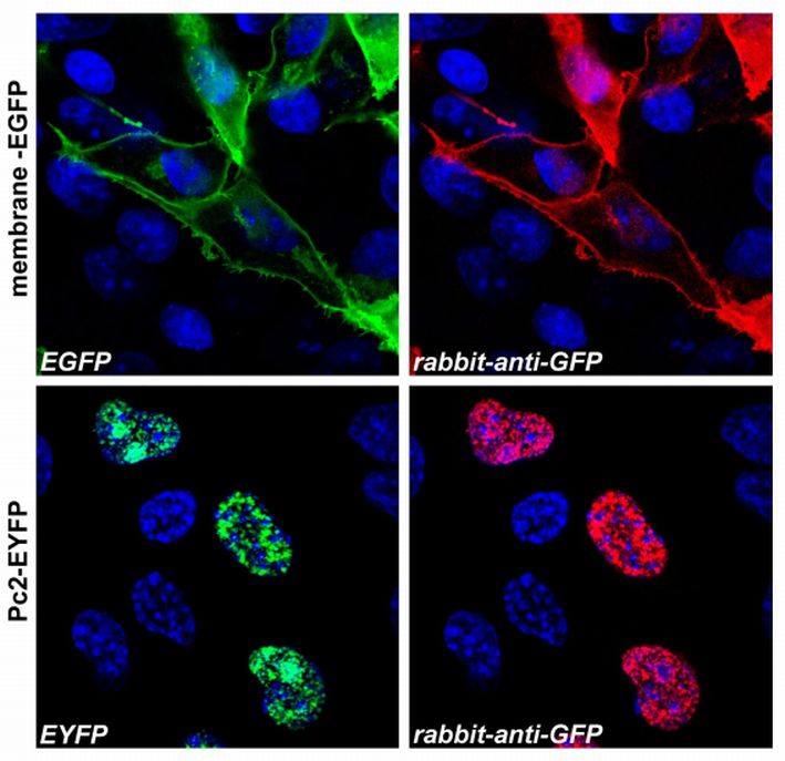
ARG62993 anti-GFP antibody ICC/IF image
Immunofluorescence: Confocal microscopy images of COS-7 cells transfected with expression constructs encoding membrane-tethered EGFP (membrane-EGFP; top) or nuclear Polycomb 2-EYFP fusion protein (Pc2-EYFP; bottom).
The natural fluorescence of the produced proteins is shown in the green channel (left), ARG62993 anti-GFP antibody signal was detected in the red channel (right). The blue nuclear stain is also shown. -
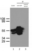
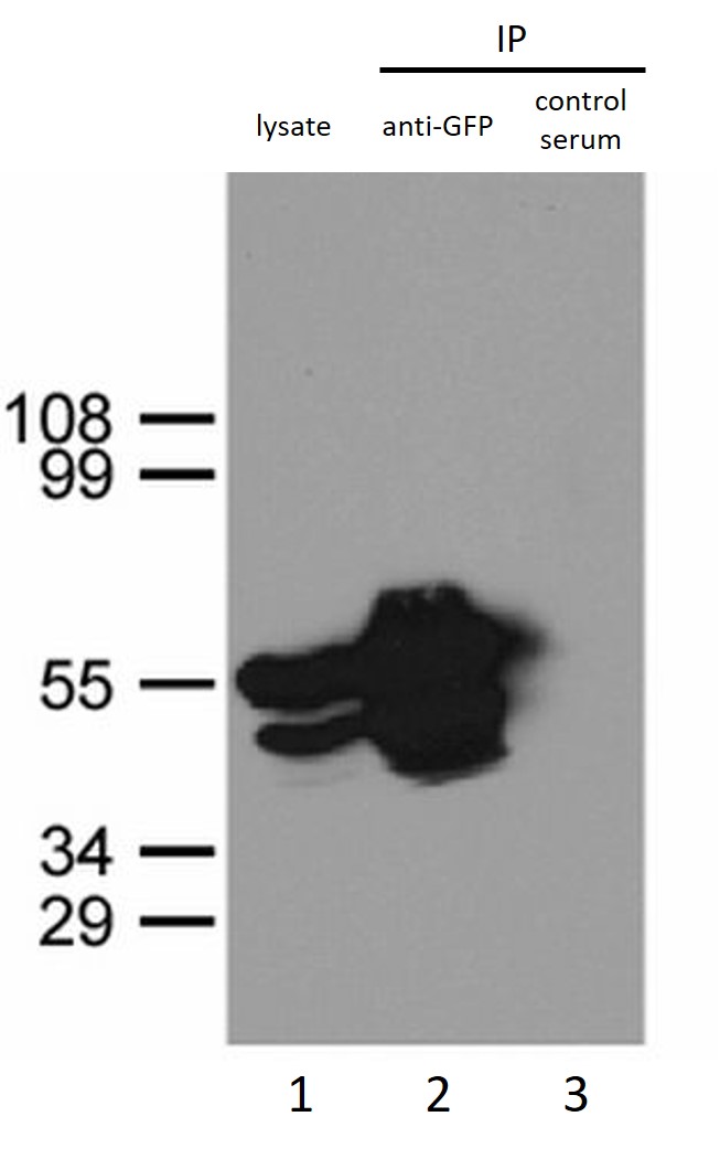
ARG62993 anti-GFP antibody IP image
Immunoprecipitation of GFP-NLS from HEK293 cells using anti-GFP antibody. HEK293 cells were transfected with expression construct encoding GFP-NLS protein. 20 hours post transfection cells were lysed in non-denaturating conditions (Lysis buffer: 20 mM Tris, pH 7.5, 100 mM NaCl, 0.5% Triton X-100, inhibitors of proteases). Aliquots of cell lysate were immunoprecipitated with ARG62993 anti-GFP antibody (lane 2) or a pre-immune rabbit serum (lane 3). Immunoprecipitates together with a sample of the cell lysate (lane 1) were separated on SDS-PAGE polyacrylamide gel and stained with ARG62993 anti-GFP antibody.
文献引用
HSPA13 modulates type I interferon antiviral pathway and NLRP3 inflammasome to restrict dengue virus infection in macrophages
WB / Human


