ARG63863
anti-GATA1 antibody
anti-GATA1 antibody for Flow cytometry,ICC/IF,Western blot and Human,Mouse
Developmental Biology antibody; Gene Regulation antibody
概述
| 产品描述 | Goat Polyclonal antibody recognizes GATA1 |
|---|---|
| 反应物种 | Hu, Ms |
| 预测物种 | Cow, Rat, Dog |
| 应用 | FACS, ICC/IF, WB |
| 宿主 | Goat |
| 克隆 | Polyclonal |
| 同位型 | IgG |
| 靶点名称 | GATA1 |
| 抗原物种 | Human |
| 抗原 | C-DAEAYRHSPVFQ |
| 偶联标记 | Un-conjugated |
| 別名 | XLTDA; Eryf1; GATA-1; GF-1; GF1; NF-E1; ERYF1; XLANP; NFE1; GATA-binding factor 1; XLTT; NF-E1 DNA-binding protein; Erythroid transcription factor |
应用说明
| 应用建议 |
|
||||||||
|---|---|---|---|---|---|---|---|---|---|
| 应用说明 | WB: Recommend incubate at RT for 1h. * The dilutions indicate recommended starting dilutions and the optimal dilutions or concentrations should be determined by the scientist. |
属性
| 形式 | Liquid |
|---|---|
| 纯化 | Purified from goat serum by antigen affinity chromatography. |
| 缓冲液 | Tris saline (pH 7.3), 0.02% Sodium azide and 0.5% BSA. |
| 抗菌剂 | 0.02% Sodium azide |
| 稳定剂 | 0.5% BSA |
| 浓度 | 0.5 mg/ml |
| 存放说明 | For continuous use, store undiluted antibody at 2-8°C for up to a week. For long-term storage, aliquot and store at -20°C or below. Storage in frost free freezers is not recommended. Avoid repeated freeze/thaw cycles. Suggest spin the vial prior to opening. The antibody solution should be gently mixed before use. |
| 注意事项 | For laboratory research only, not for drug, diagnostic or other use. |
生物信息
| 数据库连接 | |
|---|---|
| 背景介绍 | This gene encodes a protein which belongs to the GATA family of transcription factors. The protein plays an important role in erythroid development by regulating the switch of fetal hemoglobin to adult hemoglobin. Mutations in this gene have been associated with X-linked dyserythropoietic anemia and thrombocytopenia. [provided by RefSeq, Jul 2008] |
| 研究领域 | Developmental Biology antibody; Gene Regulation antibody |
| 预测分子量 | 43 kDa |
| 翻译后修饰 | Highly phosphorylated on serine residues. Phosphorylation on Ser-310 is enhanced on erythroid differentiation. Phosphorylation on Ser-142 promotes sumoylation on Lys-137 (By similarity). Sumoylation on Lys-137 is enhanced by phosphorylation on Ser-142 and by interaction with PIAS4. Sumoylation with SUMO1 has no effect on transcriptional activity (By similarity). Acetylated at 2 conserved lysine-rich motifs by CREBBP in vitro. Acetylation does not affect DNA-binding in vitro but is essential to induce erythroid differentiation and for binding chromatin in vivo (By similarity). Acetylated on Lys-233, Lys-245 Lys-246 by EP300. |
检测图片 (5) Click the Picture to Zoom In
-

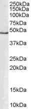
ARG63863 anti-GATA1 antibody WB image
Western Blot: human PBMC lysate (35 µg protein in RIPA buffer) stained with ARG63863 anti-GATA1 antibody at 0.3 µg/ml dilution.
-

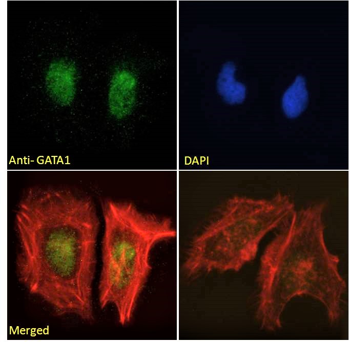
ARG63863 anti-GATA1 antibody ICC/IF image
Immunofluorescence: Paraformaldehyde fixed HeLa cells permeabilized with 0.15% Triton. Cells were stained with ARG63863 anti-GATA1 antibody (green) at 10 µg/ml dilution for 1 hour. DAPI (blue) for nuclear staining. Phalloidin (red) for Actin filaments staining. Negative control: Unimmunized goat IgG (green) at 10 µg/ml dilution.
-
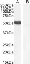
ARG63863 anti-GATA1 antibody WB image
Western blot: 35 µg of K562 nuclear lysate (A) and Human hippocampus (B, negative control) lysates (in RIPA buffer) stained with ARG63863 anti-GATA1 antibody at 1 µg/ml dilution and incubated at RT for 1 hour.
-
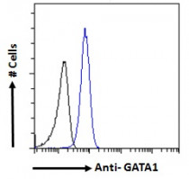
ARG63863 anti-GATA1 antibody FACS image
Flow Cytometry: Paraformaldehyde-fixed K562 cells permeabilized with 0.5% Triton. Cells were stained with ARG63863 anti-GATA1 antibody (blue line) at 10 µg/ml dilution for 1 hour, followed by incubation with Alexa FluorR 488 labelled secondary antibody. IgG control: Unimmunized goat IgG (black line).
-

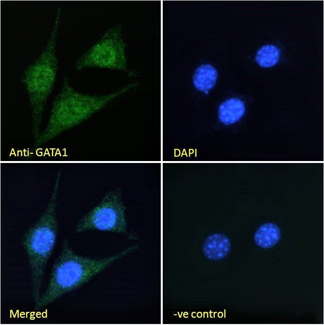
ARG63863 anti-GATA1 antibody ICC/IF image
Immunofluorescence: Paraformaldehyde fixed NIH/3T3 cells permeabilized with 0.15% Triton. Cells were stained with ARG63863 anti-GATA1 antibody (green) at 10 µg/ml dilution for 1 hour. DAPI (blue) for nuclear staining. Negative control: Unimmunized goat IgG (green) at 10 µg/ml dilution.