ARG41274
anti-ERVFRD 1 / Syncytin 2 antibody
anti-ERVFRD 1 / Syncytin 2 antibody for IHC-Formalin-fixed paraffin-embedded sections,Western blot and Human
概述
| 产品描述 | Rabbit Polyclonal antibody recognizes ERVFRD 1 / Syncytin 2 |
|---|---|
| 反应物种 | Hu |
| 应用 | IHC-P, WB |
| 宿主 | Rabbit |
| 克隆 | Polyclonal |
| 同位型 | IgG |
| 靶点名称 | ERVFRD 1 / Syncytin 2 |
| 抗原物种 | Human |
| 抗原 | Recombinant protein corresponding to aa. 16-250 of Human Syncytin 2. |
| 偶联标记 | Un-conjugated |
| 別名 | Syncytin-2; HERV-FRD; envFRD; ERVFRDE1; HERV-W/FRD; GLLL6191; Endogenous retrovirus group FRD member 1; TM; SU; Envelope polyprotein; UNQ6191; HERV-FRD_6p24.1 provirus ancestral Env polyprotein |
应用说明
| 应用建议 |
|
||||||
|---|---|---|---|---|---|---|---|
| 应用说明 | * The dilutions indicate recommended starting dilutions and the optimal dilutions or concentrations should be determined by the scientist. | ||||||
| 实际分子量 | 60-70 kDa |
属性
| 形式 | Liquid |
|---|---|
| 纯化 | Affinity purification with immunogen. |
| 缓冲液 | PBS (pH 7.3), 0.02% Sodium azide and 50% Glycerol. |
| 抗菌剂 | 0.02% Sodium azide |
| 稳定剂 | 50% Glycerol |
| 存放说明 | For continuous use, store undiluted antibody at 2-8°C for up to a week. For long-term storage, aliquot and store at -20°C. Storage in frost free freezers is not recommended. Avoid repeated freeze/thaw cycles. Suggest spin the vial prior to opening. The antibody solution should be gently mixed before use. |
| 注意事项 | For laboratory research only, not for drug, diagnostic or other use. |
生物信息
| 数据库连接 | |
|---|---|
| 基因名称 | ERVFRD-1 |
| 全名 | endogenous retrovirus group FRD, member 1 |
| 背景介绍 | Many different human endogenous retrovirus (HERV) families are expressed in normal placental tissue at high levels, suggesting that HERVs are functionally important in reproduction. This gene is part of a human endogenous retrovirus provirus on chromosome 6 that has inactivating mutations in the gag and pol genes. This gene is the envelope glycoprotein gene which appears to have been selectively preserved. The gene's protein product plays a major role in placental development and trophoblast fusion. The protein has the characteristics of a typical retroviral envelope protein, including a cleavage site that separates the surface (SU) and transmembrane (TM) proteins which form a heterodimer. [provided by RefSeq, Jun 2012] |
| 生物功能 | This endogenous retroviral envelope protein has retained its original fusogenic properties and participates in trophoblast fusion and the formation of a syncytium during placenta morphogenesis. The interaction with MFSD2A is apparently important for this process. Endogenous envelope proteins may have kept, lost or modified their original function during evolution but this one can still make pseudotypes with MLV, HIV-1 or SIV-1 virions and confer infectivity. Retroviral envelope proteins mediate receptor recognition and membrane fusion during early infection. The surface protein mediates receptor recognition, while the transmembrane protein anchors the envelope heterodimer to the viral membrane through one transmembrane domain. The other hydrophobic domain, called fusion peptide, mediates fusion of the viral membrane with the target cell membrane. [UniProt] |
| 细胞定位 | Virion. Surface protein: Cell membrane; Peripheral membrane protein. Note=The surface protein is not anchored to the membrane, but localizes to the extracellular surface through its binding to TM. Transmembrane protein: Cell membrane; Single-pass membrane protein. [UniProt] |
| 预测分子量 | 60 kDa |
| 翻译后修饰 | Specific enzymatic cleavages in vivo yield the mature SU and TM proteins. The CXXC motif is highly conserved across a broad range of retroviral envelope proteins. It is thought to participate in the formation of a labile disulfide bond possibly with the CX6CC motif present in the transmembrane protein. Isomerization of the intersubunit disulfide bond to an SU intrachain disulfide bond is thought to occur upon receptor recognition in order to allow membrane fusion (By similarity). [UniProt] |
检测图片 (2) Click the Picture to Zoom In
-
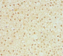
ARG41274 anti-ERVFRD 1 / Syncytin 2 antibody IHC-P image
Immunohistochemistry: Paraffin-embedded Human adrenal gland tissue stained with ARG41274 anti-ERVFRD 1 / Syncytin 2 antibody at 1:100 dilution.
-
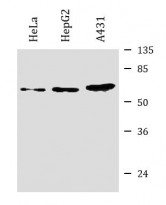
ARG41274 anti-ERVFRD 1 / Syncytin 2 antibody WB image
Western blot: HeLa, HepG2 and A431 whole cell lysates stained with ARG41274 anti-ERVFRD 1 / Syncytin 2 antibody at 4 µg/ml dilution.


