ARG43754
anti-EEA1 antibody
anti-EEA1 antibody for Flow cytometry,ICC/IF,Western blot and Human,Mouse,Rat
概述
| 产品描述 | Rabbit Polyclonal antibody recognizes EEA1 |
|---|---|
| 反应物种 | Hu, Ms, Rat |
| 应用 | FACS, ICC/IF, WB |
| 宿主 | Rabbit |
| 克隆 | Polyclonal |
| 同位型 | IgG |
| 靶点名称 | EEA1 |
| 抗原物种 | Human |
| 抗原 | Synthetic peptide corresponding to Human EEA1. |
| 偶联标记 | Un-conjugated |
| 別名 | Early endosome antigen 1; Zinc finger FYVE domain-containing protein 2; MST105; MSTP105; ZFYVE2; Endosome-associated protein p162 |
应用说明
| 应用建议 |
|
||||||||
|---|---|---|---|---|---|---|---|---|---|
| 应用说明 | * The dilutions indicate recommended starting dilutions and the optimal dilutions or concentrations should be determined by the scientist. | ||||||||
| 阳性对照 | HeLa, Placenta, Brain, Spleen, A431 | ||||||||
| 实际分子量 | ~170 kDa |
属性
| 形式 | Liquid |
|---|---|
| 纯化 | Affinity purification with immunogen. |
| 缓冲液 | 0.9% NaCl, 0.2% Na2HPO4 and 0.05% Sodium azide |
| 抗菌剂 | 0.05% Sodium azide |
| 浓度 | 0.5 mg/ml |
| 存放说明 | For continuous use, store undiluted antibody at 2-8°C for up to a week. For long-term storage, aliquot and store at -20°C. Storage in frost free freezers is not recommended. Avoid repeated freeze/thaw cycles. Suggest spin the vial prior to opening. The antibody solution should be gently mixed before use. |
| 注意事项 | For laboratory research only, not for drug, diagnostic or other use. |
生物信息
| 数据库连接 | |
|---|---|
| 基因名称 | EEA1 |
| 全名 | early endosome antigen 1 |
| 背景介绍 | Enables 1-phosphatidylinositol binding activity; GTP-dependent protein binding activity; and protein homodimerization activity. Involved in endocytosis; vesicle fusion; and viral RNA genome replication. Located in cytosol and early endosome. Is extrinsic component of plasma membrane. [provided by Alliance of Genome Resources, Apr 2022] |
| 生物功能 | Binds phospholipid vesicles containing phosphatidylinositol 3-phosphate and participates in endosomal trafficking. [UniProt] |
| 细胞定位 | Cytoplasm. Early endosome membrane; Peripheral membrane protein. [UniProt] |
| 预测分子量 | 162 kDa |
检测图片 (3) Click the Picture to Zoom In
-
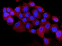
ARG43754 anti-EEA1 antibody ICC/IF image
Immunofluorescence: A431 cells were stained with ARG43754 anti-EEA1 antibody antibody (red) at 1:100 dilution at 4°C for overnight. DAPI (blue) was used for nuclear staining.
-
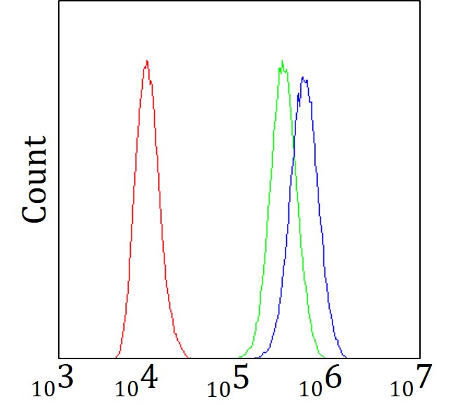
ARG43754 anti-EEA1 antibody FACS image
Flow Cytometry: A431 cells were blocked with 10% normal goat serum and stained with ARG43754 anti-EEA1 antibody (blue) at 1 µg/10^6 cells for 30 min at 20°C, followed by incubation with DyLight®488 labelled secondary antibody. Isotype control antibody (green) was rabbit IgG (1 µg/10^6 cells) used under the same conditions. Unlabelled sample (red) was also used as a control.
-
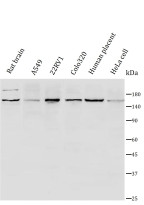
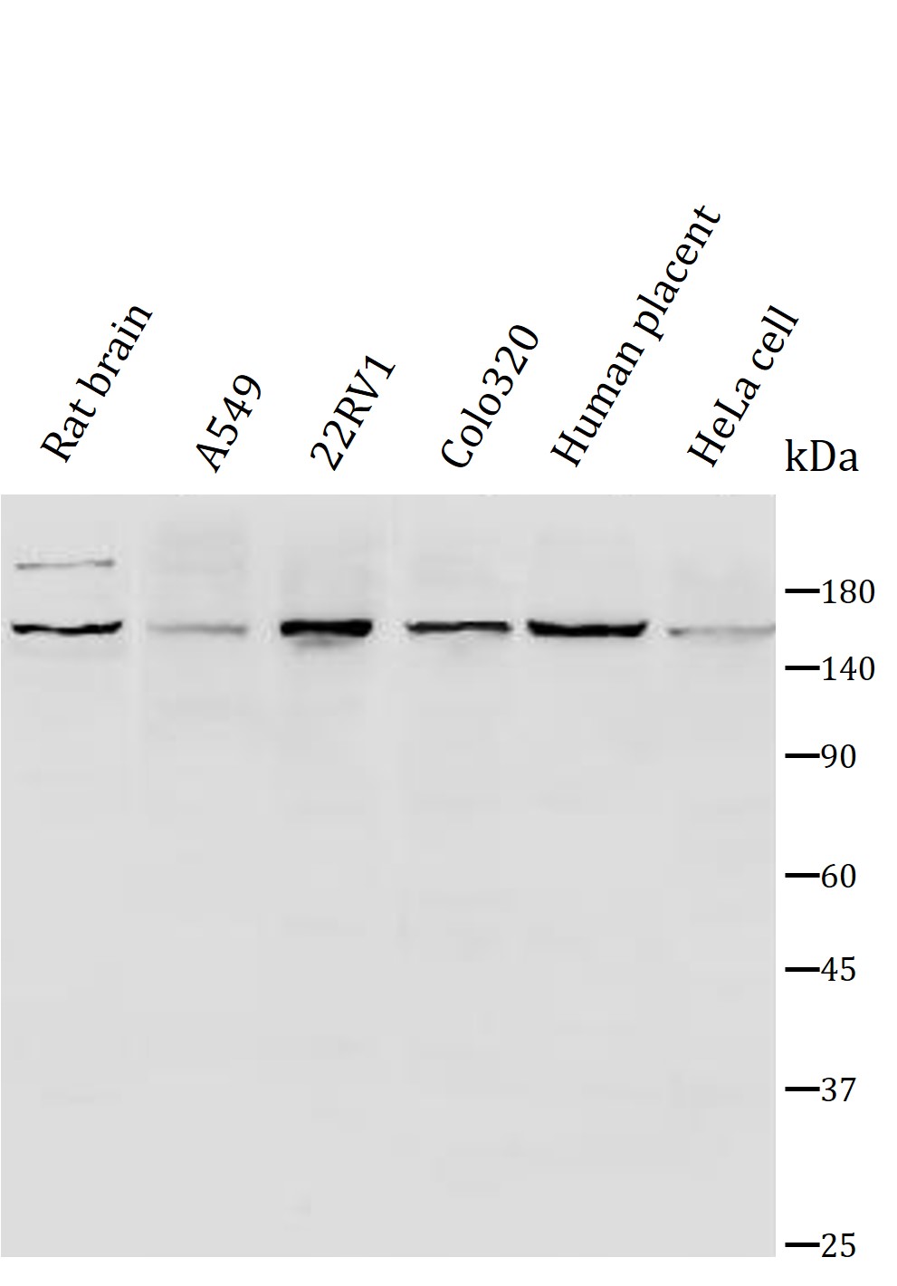
ARG43754 anti-EEA1 antibody WB image
Western blot: 1) Rat brain tissue lysates, 2) Human A549 cell lysates, 3) Human 22RV1 cell lysates, 4) Human Colo320 cell lysates, 5) Human placenta tissue lysates, 6) HeLa cell lysates, stained with ARG43754 anti-EEA1 antibody at 1:1000 dilution, overnight at 4°C.