ARG55685
anti-EBP1 antibody
anti-EBP1 antibody for Flow cytometry,IHC-Formalin-fixed paraffin-embedded sections,Western blot and Human,Mouse
概述
| 产品描述 | Rabbit Polyclonal antibody recognizes EBP1 |
|---|---|
| 反应物种 | Hu, Ms |
| 应用 | FACS, IHC-P, WB |
| 宿主 | Rabbit |
| 克隆 | Polyclonal |
| 同位型 | IgG |
| 靶点名称 | EBP1 |
| 抗原物种 | Human |
| 抗原 | KLH-conjugated synthetic peptide corresponding to aa. 228-255 (Center) of Human EBP1. |
| 偶联标记 | Un-conjugated |
| 別名 | HG4-1; p38-2G4; Proliferation-associated protein 2G4; EBP1; ErbB3-binding protein 1; hG4-1; Cell cycle protein p38-2G4 homolog |
应用说明
| 应用建议 |
|
||||||||
|---|---|---|---|---|---|---|---|---|---|
| 应用说明 | * The dilutions indicate recommended starting dilutions and the optimal dilutions or concentrations should be determined by the scientist. | ||||||||
| 阳性对照 | A2058 |
属性
| 形式 | Liquid |
|---|---|
| 纯化 | This antibody is prepared by Saturated Ammonium Sulfate (SAS) precipitation followed by dialysis against PBS. |
| 缓冲液 | PBS and 0.09% (W/V) Sodium azide. |
| 抗菌剂 | 0.09% (W/V) Sodium azide. |
| 存放说明 | For continuous use, store undiluted antibody at 2-8°C for up to a week. For long-term storage, aliquot and store at -20°C or below. Storage in frost free freezers is not recommended. Avoid repeated freeze/thaw cycles. Suggest spin the vial prior to opening. The antibody solution should be gently mixed before use. |
| 注意事项 | For laboratory research only, not for drug, diagnostic or other use. |
生物信息
| 数据库连接 |
Swiss-port # P50580 Mouse Proliferation-associated protein 2G4 Swiss-port # Q9UQ80 Human Proliferation-associated protein 2G4 |
|---|---|
| 基因名称 | PA2G4 |
| 全名 | proliferation-associated 2G4, 38kDa |
| 背景介绍 | This gene encodes an RNA-binding protein that is involved in growth regulation. This protein is present in pre-ribosomal ribonucleoprotein complexes and may be involved in ribosome assembly and the regulation of intermediate and late steps of rRNA processing. This protein can interact with the cytoplasmic domain of the ErbB3 receptor and may contribute to transducing growth regulatory signals. This protein is also a transcriptional co-repressor of androgen receptor-regulated genes and other cell cycle regulatory genes through its interactions with histone deacetylases. This protein has been implicated in growth inhibition and the induction of differentiation of human cancer cells. Six pseudogenes, located on chromosomes 3, 6, 9, 18, 20 and X, have been identified. [provided by RefSeq, Jul 2008] |
| 生物功能 | May play a role in a ERBB3-regulated signal transduction pathway. Seems be involved in growth regulation. Acts a corepressor of the androgen receptor (AR) and is regulated by the ERBB3 ligand neuregulin-1/heregulin (HRG). Inhibits transcription of some E2F1-regulated promoters, probably by recruiting histone acetylase (HAT) activity. Binds RNA. Associates with 28S, 18S and 5.8S mature rRNAs, several rRNA precursors and probably U3 small nucleolar RNA. May be involved in regulation of intermediate and late steps of rRNA processing. May be involved in ribosome assembly. Mediates cap-independent translation of specific viral IRESs (internal ribosomal entry site) (By similarity). Regulates cell proliferation, differentiation, and survival. Isoform 1 suppresses apoptosis whereas isoform 2 promotes cell differentiation (By similarity). [UniProt] |
| 细胞定位 | Isoform 1: Cytoplasm Nucleus, nucleolus. Note=Tranlocates to the nucleus upon treatment with HRG. Phosphorylation at Ser-361 by PKC/PRKCD regulates its nucleolar localization |
| 预测分子量 | 44 kDa |
| 翻译后修饰 | Phosphorylated on serine and threonine residues. Phosphorylation is enhanced by HRG treatment. Basal phosphorylation is PKC-dependent and HRG-induced phosphorylation is predominantly PKC-independent. Phosphorylation at Ser-361 by PKC/PRKCD regulates its nucleolar localization. In cancer cells, isoform 2 is polyubiquitinated leading to its proteasomal degradation and phosphorylation by PKC/PRKCD enhances polyubiquitination. |
检测图片 (3) Click the Picture to Zoom In
-
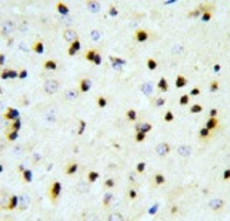
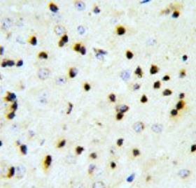
ARG55685 anti-EBP1 antibody IHC-P image
Immunohistochemistry: Formalin-fixed and paraffin-embedded Mouse brain tissue stained with ARG55685 anti-EBP1 antibody.
-
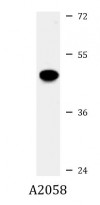
ARG55685 anti-EBP1 antibody WB image
Western blot: 35 µg of A2058 cell lysate stained with ARG55685 anti-EBP1 antibody.
-
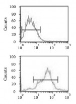
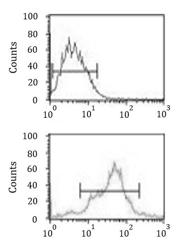
ARG55685 anti-EBP1 antibody FACS image
Flow Cytometry: MCF-7 cells stained with ARG55685 anti-EBP1 antibody (bottom histogram) or without primary antibody as control (top histogram), followed by incubation with FITC labelled secondary antibody.