ARG43367
anti-DNM1L / DRP1 antibody
anti-DNM1L / DRP1 antibody for Flow cytometry,ICC/IF,IHC-Formalin-fixed paraffin-embedded sections,Western blot and Human,Mouse,Rat
概述
| 产品描述 | Rabbit Polyclonal antibody recognizes DNM1L / DRP1 |
|---|---|
| 反应物种 | Hu, Ms, Rat |
| 应用 | FACS, ICC/IF, IHC-P, WB |
| 宿主 | Rabbit |
| 克隆 | Polyclonal |
| 同位型 | IgG |
| 靶点名称 | DNM1L / DRP1 |
| 抗原物种 | Human |
| 抗原 | Recombinant protein corresponding to Q618-W736 of Human DNM1L / DRP1. |
| 偶联标记 | Un-conjugated |
| 別名 | EMPF; Dynamin family member proline-rich carboxyl-terminal domain less; Dynamin-1-like protein; DLP1; HDYNIV; Dynamin-like protein 4; DRP1; Dynamin-related protein 1; Dynamin-like protein; Dnm1p/Vps1p-like protein; EC 3.6.5.5; DVLP; HdynIV; Dymple; DYMPLE; Dynamin-like protein IV |
应用说明
| 应用建议 |
|
||||||||||
|---|---|---|---|---|---|---|---|---|---|---|---|
| 应用说明 | IHC-P: Antigen Retrieval: Heat mediation was performed in EDTA buffer (pH 8.0). * The dilutions indicate recommended starting dilutions and the optimal dilutions or concentrations should be determined by the scientist. |
||||||||||
| 实际分子量 | ~ 83 kDa |
属性
| 形式 | Liquid |
|---|---|
| 纯化 | Affinity purification with immunogen. |
| 缓冲液 | 0.2% Na2HPO4, 0.9% NaCl, 0.01% Sodium azide and 4% Trehalose. |
| 抗菌剂 | 0.01% Sodium azide |
| 稳定剂 | 4% Trehalose |
| 浓度 | 0.5 mg/ml |
| 存放说明 | For continuous use, store undiluted antibody at 2-8°C for up to a week. For long-term storage, aliquot and store at -20°C or below. Storage in frost free freezers is not recommended. Avoid repeated freeze/thaw cycles. Suggest spin the vial prior to opening. The antibody solution should be gently mixed before use. |
| 注意事项 | For laboratory research only, not for drug, diagnostic or other use. |
生物信息
| 数据库连接 | |
|---|---|
| 基因名称 | DNM1L |
| 全名 | dynamin 1-like |
| 背景介绍 | This gene encodes a member of the dynamin superfamily of GTPases. The encoded protein mediates mitochondrial and peroxisomal division, and is involved in developmentally regulated apoptosis and programmed necrosis. Dysfunction of this gene is implicated in several neurological disorders, including Alzheimer's disease. Mutations in this gene are associated with the autosomal dominant disorder, encephalopathy, lethal, due to defective mitochondrial and peroxisomal fission (EMPF). Alternative splicing results in multiple transcript variants encoding different isoforms. [provided by RefSeq, Jun 2013] |
| 生物功能 | Functions in mitochondrial and peroxisomal division. Mediates membrane fission through oligomerization into membrane-associated tubular structures that wrap around the scission site to constrict and sever the mitochondrial membrane through a GTP hydrolysis-dependent mechanism. The specific recruitment at scission sites is mediated by membrane receptors like MFF, MIEF1 and MIEF2 for mitochondrial membranes (PubMed:29899447). While the recruitment by the membrane receptors is GTP-dependent, the following hydrolysis of GTP induces the dissociation from the receptors and allows DNM1L filaments to curl into closed rings that are probably sufficient to sever a double membrane (PubMed:29899447). Through its function in mitochondrial division, ensures the survival of at least some types of postmitotic neurons, including Purkinje cells, by suppressing oxidative damage. Required for normal brain development, including that of cerebellum. Facilitates developmentally regulated apoptosis during neural tube formation. Required for a normal rate of cytochrome c release and caspase activation during apoptosis; this requirement may depend upon the cell type and the physiological apoptotic cues. Plays an important role in mitochondrial fission during mitosis (PubMed:26992161, PubMed:27301544, PubMed:27328748). Required for formation of endocytic vesicles. Proposed to regulate synaptic vesicle membrane dynamics through association with BCL2L1 isoform Bcl-X(L) which stimulates its GTPase activity in synaptic vesicles; the function may require its recruitment by MFF to clathrin-containing vesicles (PubMed:23792689). Required for programmed necrosis execution. Rhythmic control of its activity following phosphorylation at Ser-637 is essential for the circadian control of mitochondrial ATP production (PubMed:29478834). [Isoform 1]: Inhibits peroxisomal division when overexpressed. [Isoform 4]: Inhibits peroxisomal division when overexpressed. [UniProt] |
| 细胞定位 | Cytoplasm, cytosol. Golgi apparatus. Endomembrane system; Peripheral membrane protein. Mitochondrion outer membrane; Peripheral membrane protein. Peroxisome. Membrane, clathrin-coated pit. Cytoplasmic vesicle, secretory vesicle, synaptic vesicle membrane. Note=Mainly cytosolic. Translocated to the mitochondrial membrane through O-GlcNAcylation and interaction with FIS1. Recruited to the mitochondrial outer membrane by interaction with MIEF1. [UniProt] |
| 预测分子量 | 82 kDa |
| 翻译后修饰 | Phosphorylation/dephosphorylation events on two sites near the GED domain regulate mitochondrial fission. Phosphorylation on Ser-637 inhibits the GTPase activity, leading to a defect in mitochondrial fission promoting mitochondrial elongation. Dephosphorylated on this site by PPP3CA which promotes mitochondrial fission. Phosphorylation on Ser-616 activates the GTPase activity and promotes mitochondrial fission. Sumoylated on various lysine residues within the B domain, probably by MUL1. Sumoylation positively regulates mitochondrial fission. Desumoylated by SENP5 during G2/M transition of mitosis. Appears to be linked to its catalytic activity. S-nitrosylation increases DNM1L dimerization, mitochondrial fission and causes neuronal damage. Ubiquitination by MARCH5 affects mitochondrial morphology. O-GlcNAcylation augments the level of the GTP-bound active form of DRP1 and induces translocation from the cytoplasm to mitochondria in cardiomyocytes. It also decreases phosphorylation at Ser-637 (By similarity). [UniProt] |
检测图片 (13) Click the Picture to Zoom In
-

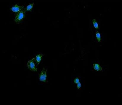
ARG43367 anti-DNM1L / DRP1 antibody ICC/IF image
Immunofluorescence: U2OS cells stained with ARG43367 anti-DNM1L / DRP1 antibody (green) at 5 µg/ml dilution, overnight at 4°C. DAPI (blue) for nuclear staining.
-

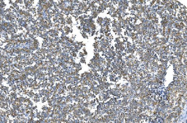
ARG43367 anti-DNM1L / DRP1 antibody IHC-P image
Immunohistochemistry: Paraffin-embedded Human tonsil tissue. Antigen Retrieval: Heat mediation was performed in EDTA buffer (pH 8.0). The tissue section was blocked with 10% goat serum. The tissue section was then stained with ARG43367 anti-DNM1L / DRP1 antibody at 2 µg/ml dilution, overnight at 4°C.
-
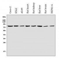
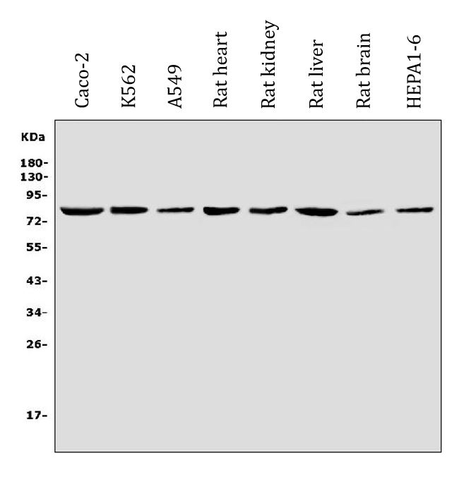
ARG43367 anti-DNM1L / DRP1 antibody WB image
Western blot: 30 µg of sample under reducing conditions. Caco-2, K562, A549, Rat heart, Rat kidney, Rat liver, Rat brain and HEPA1-6 whole cell lysates stained with ARG43367 anti-DNM1L / DRP1 antibody at 0.25 µg/ml dilution, overnight at 4°C.
-
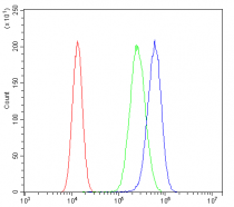
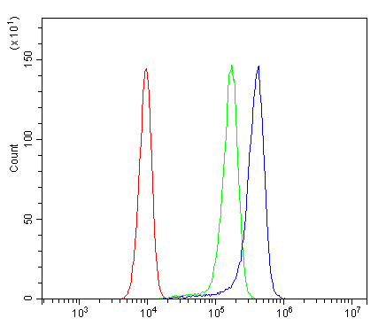
ARG43367 anti-DNM1L / DRP1 antibody FACS image
Flow Cytometry: A549 cells were blocked with 10% normal goat serum and then stained with ARG43367 anti-DNM1L / DRP1 antibody (blue) at 1 µg/10^6 cells for 30 min at 20°C, followed by incubation with DyLight®488 labelled secondary antibody. Isotype control antibody (green) was rabbit IgG (1 µg/10^6 cells) used under the same conditions. Unlabelled sample (red) was also used as a control.
-
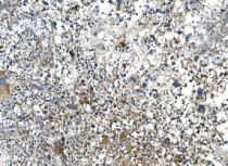
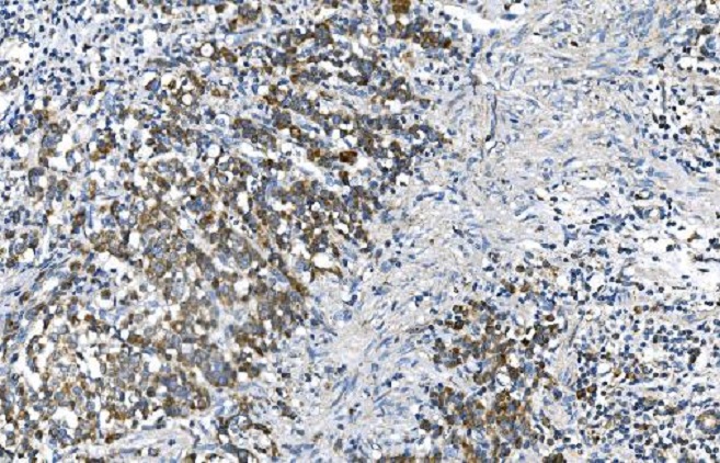
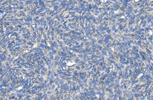
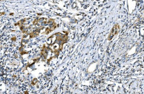
ARG43367 anti-DNM1L / DRP1 antibody IHC-P image
Immunohistochemistry: Paraffin-embedded Human liver cancer tissue. Antigen Retrieval: Heat mediation was performed in EDTA buffer (pH 8.0). The tissue section was blocked with 10% goat serum. The tissue section was then stained with ARG43367 anti-DNM1L / DRP1 antibody at 2 µg/ml dilution, overnight at 4°C.
-
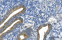
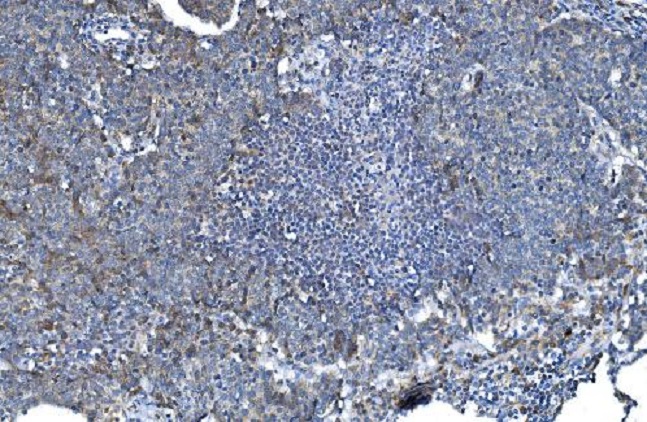
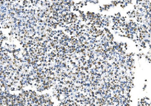
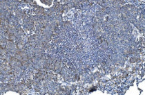
ARG43367 anti-DNM1L / DRP1 antibody IHC-P image
Immunohistochemistry: Paraffin-embedded Human appendicitis tissue. Antigen Retrieval: Heat mediation was performed in EDTA buffer (pH 8.0). The tissue section was blocked with 10% goat serum. The tissue section was then stained with ARG43367 anti-DNM1L / DRP1 antibody at 2 µg/ml dilution, overnight at 4°C.
-
ARG43367 anti-DNM1L / DRP1 antibody IHC-P image
Immunohistochemistry: Paraffin-embedded Human bladder cancer tissue. Antigen Retrieval: Heat mediation was performed in EDTA buffer (pH 8.0). The tissue section was blocked with 10% goat serum. The tissue section was then stained with ARG43367 anti-DNM1L / DRP1 antibody at 2 µg/ml dilution, overnight at 4°C.
-

ARG43367 anti-DNM1L / DRP1 antibody IHC-P image
Immunohistochemistry: Paraffin-embedded Human gastric cancer tissue. Antigen Retrieval: Heat mediation was performed in EDTA buffer (pH 8.0). The tissue section was blocked with 10% goat serum. The tissue section was then stained with ARG43367 anti-DNM1L / DRP1 antibody at 2 µg/ml dilution, overnight at 4°C.
-
ARG43367 anti-DNM1L / DRP1 antibody IHC-P image
Immunohistochemistry: Paraffin-embedded Human lung cancer tissue. Antigen Retrieval: Heat mediation was performed in EDTA buffer (pH 8.0). The tissue section was blocked with 10% goat serum. The tissue section was then stained with ARG43367 anti-DNM1L / DRP1 antibody at 2 µg/ml dilution, overnight at 4°C.
-

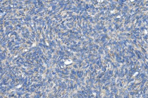
ARG43367 anti-DNM1L / DRP1 antibody IHC-P image
Immunohistochemistry: Paraffin-embedded Human melanoma tissue. Antigen Retrieval: Heat mediation was performed in EDTA buffer (pH 8.0). The tissue section was blocked with 10% goat serum. The tissue section was then stained with ARG43367 anti-DNM1L / DRP1 antibody at 2 µg/ml dilution, overnight at 4°C.
-
ARG43367 anti-DNM1L / DRP1 antibody IHC-P image
Immunohistochemistry: Paraffin-embedded Human skin cancer tissue. Antigen Retrieval: Heat mediation was performed in EDTA buffer (pH 8.0). The tissue section was blocked with 10% goat serum. The tissue section was then stained with ARG43367 anti-DNM1L / DRP1 antibody at 2 µg/ml dilution, overnight at 4°C.
-

ARG43367 anti-DNM1L / DRP1 antibody IHC-P image
Immunohistochemistry: Paraffin-embedded Human testicular cancer tissue. Antigen Retrieval: Heat mediation was performed in EDTA buffer (pH 8.0). The tissue section was blocked with 10% goat serum. The tissue section was then stained with ARG43367 anti-DNM1L / DRP1 antibody at 2 µg/ml dilution, overnight at 4°C.
-
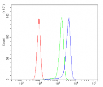
ARG43367 anti-DNM1L / DRP1 antibody FACS image
Flow Cytometry: SiHa cells were blocked with 10% normal goat serum and then stained with ARG43367 anti-DNM1L / DRP1 antibody (blue) at 1 µg/10^6 cells for 30 min at 20°C, followed by incubation with DyLight®488 labelled secondary antibody. Isotype control antibody (green) was rabbit IgG (1 µg/10^6 cells) used under the same conditions. Unlabelled sample (red) was also used as a control.