ARG44234
anti-DAPK1 antibody
anti-DAPK1 antibody for IHC-Formalin-fixed paraffin-embedded sections,Western blot and Human,Mouse,Rat
概述
| 产品描述 | Rabbit Polyclonal antibody recognizes DAPK1 |
|---|---|
| 反应物种 | Hu, Ms, Rat |
| 应用 | IHC-P, WB |
| 宿主 | Rabbit |
| 克隆 | Polyclonal |
| 同位型 | IgG |
| 靶点名称 | DAPK1 |
| 抗原物种 | Human |
| 抗原 | Recombinant protein of Human DAPK1 |
| 偶联标记 | Un-conjugated |
| Protein Full name | Death-associated protein kinase 1 |
| 別名 | DAPK1; Death Associated Protein Kinase 1; DAPK; Death-Associated Protein Kinase 1; DAP Kinase 1; EC 2.7.11.1; EC 2.7.11; ROCO3 |
应用说明
| 应用建议 |
|
||||||
|---|---|---|---|---|---|---|---|
| 应用说明 | * The dilutions indicate recommended starting dilutions and the optimal dilutions or concentrations should be determined by the scientist. |
属性
| 形式 | Liquid |
|---|---|
| 纯化 | Affinity purification with immunogen. |
| 缓冲液 | 0.9% NaCl, 0.2% Na2HPO4, 0.05% Sodium azide and 5% BSA. |
| 抗菌剂 | 0.05% Sodium azide |
| 稳定剂 | 5% BSA |
| 浓度 | 0.5 mg/ml |
| 存放说明 | For continuous use, store undiluted antibody at 2-8°C for up to a week. For long-term storage, aliquot and store at -20°C or below. Storage in frost free freezers is not recommended. Avoid repeated freeze/thaw cycles. Suggest spin the vial prior to opening. The antibody solution should be gently mixed before use. |
| 注意事项 | For laboratory research only, not for drug, diagnostic or other use. |
生物信息
| 数据库连接 | |
|---|---|
| 基因名称 | DAPK1 |
| 全名 | Death Associated Protein Kinase 1 |
| 背景介绍 | Death-associated protein kinase 1 is a positive mediator of gamma-interferon induced programmed cell death. DAPK1 encodes a structurally unique 160-kD calmodulin dependent serine-threonine kinase that carries 8 ankyrin repeats and 2 putative P-loop consensus sites. It is a tumor suppressor candidate. Alternative splicing results in multiple transcript variants. |
| 生物功能 | Calcium/calmodulin-dependent serine/threonine kinase involved in multiple cellular signaling pathways that trigger cell survival, apoptosis, and autophagy. Regulates both type I apoptotic and type II autophagic cell deaths signal, depending on the cellular setting. The former is caspase-dependent, while the latter is caspase-independent and is characterized by the accumulation of autophagic vesicles. Phosphorylates PIN1 resulting in inhibition of its catalytic activity, nuclear localization, and cellular function. Phosphorylates TPM1, enhancing stress fiber formation in endothelial cells. Phosphorylates STX1A and significantly decreases its binding to STXBP1. Phosphorylates PRKD1 and regulates JNK signaling by binding and activating PRKD1 under oxidative stress. Phosphorylates BECN1, reducing its interaction with BCL2 and BCL2L1 and promoting the induction of autophagy. Phosphorylates TSC2, disrupting the TSC1-TSC2 complex and stimulating mTORC1 activity in a growth factor-dependent pathway. Phosphorylates RPS6, MYL9 and DAPK3. Acts as a signaling amplifier of NMDA receptors at extrasynaptic sites for mediating brain damage in stroke. Cerebral ischemia recruits DAPK1 into the NMDA receptor complex and it phosphorylates GRINB at Ser-1303 inducing injurious Ca2+ influx through NMDA receptor channels, resulting in an irreversible neuronal death. Required together with DAPK3 for phosphorylation of RPL13A upon interferon-gamma activation which is causing RPL13A involvement in transcript-selective translation inhibition. |
| 细胞定位 | Cytoplasm, Cytoskeleton |
| 预测分子量 | 160 kDa |
| 翻译后修饰 | Phosphoprotein, Ubl conjugation |
检测图片 (4) Click the Picture to Zoom In
-
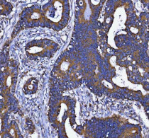
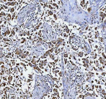
ARG44234 anti-DAPK1 antibody IHC-P image
Immunohistochemistry: Human colorectal adenocarcinoma stained with ARG44234 anti-DAPK1 antibody at 2 μg/ml dilution.
-
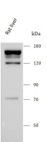
ARG44234 anti-DAPK1 antibody WB image
Western blot: Rat liver stained with ARG44234 anti-DAPK1 antibody at 0.5 μg/mL dilution.
-
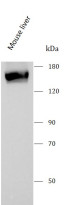
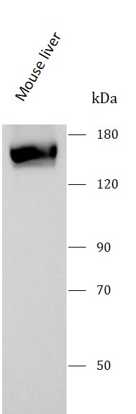
ARG44234 anti-DAPK1 antibody WB image
Western blot: Mouse liver stained with ARG44234 anti-DAPK1 antibody at 0.5 μg/mL dilution.
-
ARG44234 anti-DAPK1 antibody IHC-P image
Immunohistochemistry: Human lung cancer stained with ARG44234 anti-DAPK1 antibody at 2 μg/ml dilution.