ARG63209
anti-DAP3 antibody
anti-DAP3 antibody for ICC/IF,IHC-Formalin-fixed paraffin-embedded sections,Western blot and Human
Cell Biology and Cellular Response antibody; Cell Death antibody; Gene Regulation antibody; Metabolism antibody
概述
| 产品描述 | Goat Polyclonal antibody recognizes DAP3 |
|---|---|
| 反应物种 | Hu |
| 应用 | ICC/IF, IHC-P, WB |
| 特异性 | This antibody is expected to recognise isoform 1 (NP_387506.1), isoform 2 (NP_001186779.1) and isoform 3 (NP_001186780.1). Reported variants represent identical protein (NP_387506.1; NP_004623.1; NP_001186778.1). |
| 宿主 | Goat |
| 克隆 | Polyclonal |
| 同位型 | IgG |
| 靶点名称 | DAP3 |
| 抗原物种 | Human |
| 抗原 | NPSLLERHCAYL |
| 偶联标记 | Un-conjugated |
| 別名 | Ionizing radiation resistance conferring protein; 28S ribosomal protein S29, mitochondrial; Death-associated protein 3; MRPS29; bMRP-10; DAP-3; S29mt; MRP-S29 |
应用说明
| 应用建议 |
|
||||||||
|---|---|---|---|---|---|---|---|---|---|
| 应用说明 | WB: Recommend incubate at RT for 1h. IHC-P: Antigen Retrieval: Steam tissue section in Citrate buffer (pH 6.0). * The dilutions indicate recommended starting dilutions and the optimal dilutions or concentrations should be determined by the scientist. |
属性
| 形式 | Liquid |
|---|---|
| 纯化 | Purified from goat serum by antigen affinity chromatography. |
| 缓冲液 | Tris saline (pH 7.3), 0.02% Sodium azide and 0.5% BSA. |
| 抗菌剂 | 0.02% Sodium azide |
| 稳定剂 | 0.5% BSA |
| 浓度 | 0.5 mg/ml |
| 存放说明 | For continuous use, store undiluted antibody at 2-8°C for up to a week. For long-term storage, aliquot and store at -20°C or below. Storage in frost free freezers is not recommended. Avoid repeated freeze/thaw cycles. Suggest spin the vial prior to opening. The antibody solution should be gently mixed before use. |
| 注意事项 | For laboratory research only, not for drug, diagnostic or other use. |
生物信息
| 数据库连接 |
Swiss-port # P51398 Human 28S ribosomal protein S29, mitochondrial |
|---|---|
| 背景介绍 | Mammalian mitochondrial ribosomal proteins are encoded by nuclear genes and help in protein synthesis within the mitochondrion. Mitochondrial ribosomes (mitoribosomes) consist of a small 28S subunit and a large 39S subunit. They have an estimated 75% protein to rRNA composition compared to prokaryotic ribosomes, where this ratio is reversed. Another difference between mammalian mitoribosomes and prokaryotic ribosomes is that the latter contain a 5S rRNA. Among different species, the proteins comprising the mitoribosome differ greatly in sequence, and sometimes in biochemical properties, which prevents easy recognition by sequence homology. This gene encodes a 28S subunit protein that also participates in apoptotic pathways which are initiated by tumor necrosis factor-alpha, Fas ligand, and gamma interferon. This protein potentially binds ATP/GTP and might be a functional partner of the mitoribosomal protein S27. Multiple alternatively spliced transcript variants encoding distinct isoforms have been found for this gene. Pseudogenes corresponding to this gene are found on chromosomes 1q and 2q. [provided by RefSeq, Dec 2010] |
| 研究领域 | Cell Biology and Cellular Response antibody; Cell Death antibody; Gene Regulation antibody; Metabolism antibody |
| 预测分子量 | 46 kDa |
检测图片 (8) Click the Picture to Zoom In
-
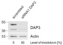
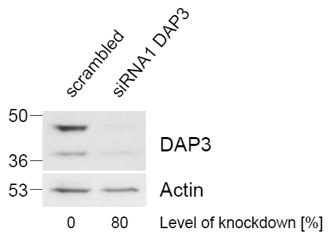
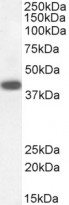
ARG63209 anti-DAP3 antibody WB image
Western Blot: HeLa lysate (control in left lane and after si-RNA-mediated DAP3 knock-down expresson in right lane) (35 µg protein in RIPA buffer) Level of knock-down relative to Actin expression level was determined by RT-PCR. stained with ARG63209 anti-DAP3 antibody at 1 µg/ml dilution.
-

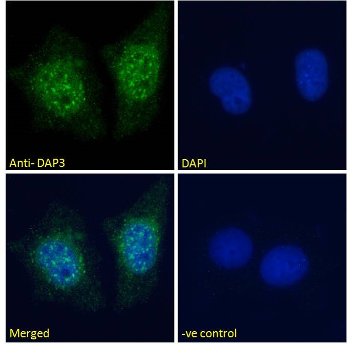
ARG63209 anti-DAP3 antibody ICC/IF image
Immunofluorescence: Paraformaldehyde fixed MCF7 cells permeabilized with 0.15% Triton. Cells were stained with ARG63209 anti-DAP3 antibody (green) at 10 µg/ml dilution for 1 hour. DAPI (blue) for nuclear staining. Negative control: Unimmunized goat IgG (green) at 10 µg/ml dilution.
-
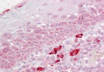
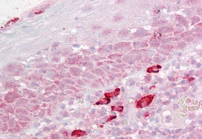
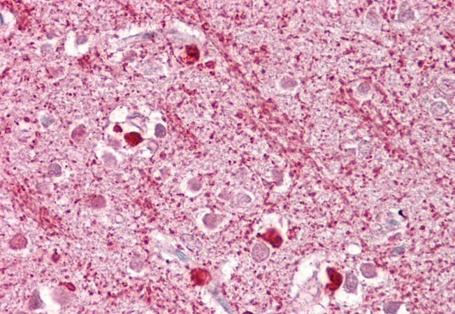
ARG63209 anti-DAP3 antibody IHC-P image
Immunohistochemistry: Paraffin-embedded Human tonsil tissue. Antigen Retrieval: Steam tissue section in Citrate buffer (pH 6.0). The tissue section was stained with ARG63209 anti-DAP3 antibody at 2.5 µg/ml dilution followed by AP-staining.
-
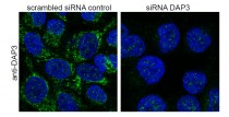
ARG63209 anti-DAP3 antibody ICC/IF image
Immunofluorescence: guanidinium thiocyanate-treated HeLa before (left) and after (right) si-RNA-mediated DAP3 knock-down expresson stained with ARG63209 anti-DAP3 antibod (0.5ug/ml). Primary incubation 1h at ambient temp. Detection by DyLight 488. Nuclear DAPI stain.
-

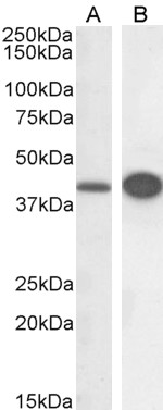
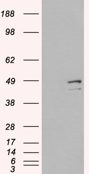
ARG63209 anti-DAP3 antibody WB image
Western blot: 30 µg of HeLa (A) and HepG2 (B) cell lysates (in RIPA buffer) stained with ARG63209 anti-DAP3 antibody at 0.3 µg/ml dilution and incubated at RT for 1 hour.
-
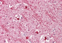
ARG63209 anti-DAP3 antibody IHC-P image
Immunohistochemistry: Paraffin-embedded Human cortex tissue. Antigen Retrieval: Steam tissue section in Citrate buffer (pH 6.0). The tissue section was stained with ARG63209 anti-DAP3 antibody at 2.5 µg/ml dilution followed by AP-staining.
-
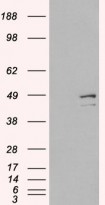
ARG63209 anti-DAP3 antibody WB image
Western Blot: 1). Mock transfection; 2) DAP3 (RC223182) expressing plasmid transfected HEK293 cell lysate standed with ARG63209 anti-DAP3 antibody
-
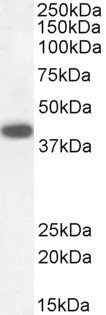
ARG63209 anti-DAP3 antibody WB image
Western blot: 30 µg of Human kidney lysate (in RIPA buffer) stained with ARG63209 anti-DAP3 antibody at 0.3 µg/ml dilution and incubated at RT for 1 hour.