ARG56953
anti-Cyclophilin B antibody [k2E2]
anti-Cyclophilin B antibody [k2E2] for Flow cytometry,Western blot and Human
概述
| 产品描述 | Mouse Monoclonal antibody [k2E2] recognizes Cyclophilin B |
|---|---|
| 反应物种 | Hu |
| 应用 | FACS, WB |
| 宿主 | Mouse |
| 克隆 | Monoclonal |
| 克隆号 | k2E2 |
| 同位型 | IgG1, kappa |
| 靶点名称 | Cyclophilin B |
| 抗原物种 | Human |
| 抗原 | Recombinant fragment around aa. 26-216 of Human Cyclophilin B. |
| 偶联标记 | Un-conjugated |
| 別名 | HEL-S-39; CYPB; PPIase B; CYP-S1; Cyclophilin B; SCYLP; EC 5.2.1.8; S-cyclophilin; OI9; Rotamase B; Peptidyl-prolyl cis-trans isomerase B |
应用说明
| 应用建议 |
|
||||||
|---|---|---|---|---|---|---|---|
| 应用说明 | * The dilutions indicate recommended starting dilutions and the optimal dilutions or concentrations should be determined by the scientist. |
属性
| 形式 | Liquid |
|---|---|
| 纯化 | Purification with Protein G. |
| 缓冲液 | PBS (pH 7.4), 0.02% Sodium azide and 10% Glycerol. |
| 抗菌剂 | 0.02% Sodium azide |
| 稳定剂 | 10% Glycerol |
| 浓度 | 1 mg/ml |
| 存放说明 | For continuous use, store undiluted antibody at 2-8°C for up to a week. For long-term storage, aliquot and store at -20°C. Storage in frost free freezers is not recommended. Avoid repeated freeze/thaw cycles. Suggest spin the vial prior to opening. The antibody solution should be gently mixed before use. |
| 注意事项 | For laboratory research only, not for drug, diagnostic or other use. |
生物信息
| 数据库连接 |
Swiss-port # P23284 Human Peptidyl-prolyl cis-trans isomerase B |
|---|---|
| 基因名称 | PPIB |
| 全名 | peptidylprolyl isomerase B (cyclophilin B) |
| 背景介绍 | The protein encoded by this gene is a cyclosporine-binding protein and is mainly located within the endoplasmic reticulum. It is associated with the secretory pathway and released in biological fluids. This protein can bind to cells derived from T- and B-lymphocytes, and may regulate cyclosporine A-mediated immunosuppression. Variants have been identified in this protein that give rise to recessive forms of osteogenesis imperfecta. [provided by RefSeq, Oct 2009] |
| 生物功能 | PPIases accelerate the folding of proteins. It catalyzes the cis-trans isomerization of proline imidic peptide bonds in oligopeptides. [UniProt] |
| 预测分子量 | 24 kDa |
检测图片 (4) Click the Picture to Zoom In
-

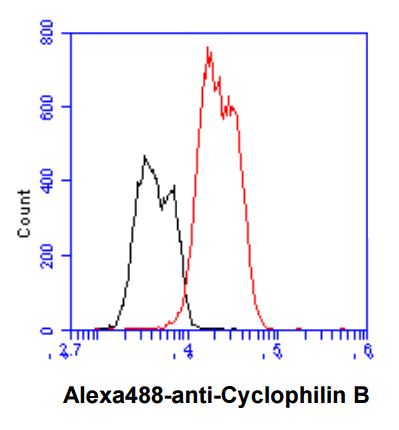
ARG56953 anti-Cyclophilin B antibody [k2E2] FACS image
Flow Cytometry: Hep3B cell line stained with ARG56953 anti-Cyclophilin B antibody [k2E2] at 2-5 µg for 1x10^6 cells (red line). Secondary antibody: Goat anti-Mouse IgG Alexa fluor 488 conjugate. Isotype control antibody was Mouse IgG (black line).
-
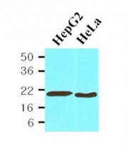
ARG56953 anti-Cyclophilin B antibody [k2E2] WB image
Western blot: 30 µg HepG2 and HeLa cell lysates stained with ARG56953 anti-Cyclophilin B antibody [k2E2] at 1:1000.
-
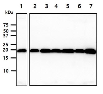
ARG56953 anti-Cyclophilin B antibody [k2E2] WB image
Western blot: 40 µg of 1) Jurkat cell lysate, 2) K562 cell lysate, 3) 293T cell lysate, 4) A549 cell lysate, 5) MCF7 cell lysate, 6) SK-OV-3 cell lysate, 7) LnCap cell lysate stained with ARG56953 anti-Cyclophilin B antibody [k2E2] at 1:1000.
-
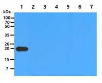
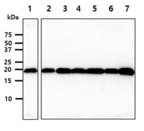
ARG56953 anti-Cyclophilin B antibody [k2E2] WB image
Western blot: 50 ng of 1) Cyclophilin B Recombinant Protein, 2) Cyclophilin A Recombinant Protein, 3) Cyclophilin D Recombinant Protein, 4) Cyclophilin E Recombinant Protein, 5) Cyclophilin F Recombinant Protein, 6) Cyclophilin G Recombinant Protein, 7) Cyclophilin H Recombinant Protein stained with ARG56953 anti-Cyclophilin B antibody [k2E2] at 1:1000.