ARG59492
anti-Collagen XVII antibody
anti-Collagen XVII antibody for Flow cytometry,Western blot and Human
概述
| 产品描述 | Rabbit Polyclonal antibody recognizes Collagen XVII |
|---|---|
| 反应物种 | Hu |
| 应用 | FACS, WB |
| 宿主 | Rabbit |
| 克隆 | Polyclonal |
| 同位型 | IgG |
| 靶点名称 | Collagen XVII |
| 抗原物种 | Human |
| 抗原 | KLH-conjugated synthetic peptide between aa. 475-504 of Human Collagen XVII. |
| 偶联标记 | Un-conjugated |
| 別名 | ERED; BP180; BPA-2; BPAG2; LAD-1; BA16H23.2; Collagen alpha-1(XVII) chain; 180 kDa bullous pemphigoid antigen 2; Bullous pemphigoid antigen 2; 120 kDa linear IgA dermatosis antigen; Linear IgA disease antigen 1; LAD-1; 97 kDa linear IgA disease antigen; 97 kDa linear IgA bullous dermatosis antigen; 97 kDa LAD antigen; 97-LAD; Linear IgA bullous disease antigen of 97 kDa; LABD97 |
应用说明
| 应用建议 |
|
||||||
|---|---|---|---|---|---|---|---|
| 应用说明 | * The dilutions indicate recommended starting dilutions and the optimal dilutions or concentrations should be determined by the scientist. | ||||||
| 阳性对照 | A2058 |
属性
| 形式 | Liquid |
|---|---|
| 纯化 | Purification with Protein A and immunogen peptide. |
| 缓冲液 | PBS and 0.09% (W/V) Sodium azide. |
| 抗菌剂 | 0.09% (W/V) Sodium azide. |
| 存放说明 | For continuous use, store undiluted antibody at 2-8°C for up to a week. For long-term storage, aliquot and store at -20°C or below. Storage in frost free freezers is not recommended. Avoid repeated freeze/thaw cycles. Suggest spin the vial prior to opening. The antibody solution should be gently mixed before use. |
| 注意事项 | For laboratory research only, not for drug, diagnostic or other use. |
生物信息
| 数据库连接 | |
|---|---|
| 基因名称 | COL17A1 |
| 全名 | collagen, type XVII, alpha 1 |
| 背景介绍 | This gene encodes the alpha chain of type XVII collagen. Unlike most collagens, collagen XVII is a transmembrane protein. Collagen XVII is a structural component of hemidesmosomes, multiprotein complexes at the dermal-epidermal basement membrane zone that mediate adhesion of keratinocytes to the underlying membrane. Mutations in this gene are associated with both generalized atrophic benign and junctional epidermolysis bullosa. Two homotrimeric forms of type XVII collagen exist. The full length form is the transmembrane protein. A soluble form, referred to as either ectodomain or LAD-1, is generated by proteolytic processing of the full length form. [provided by RefSeq, Jul 2008] |
| 生物功能 | May play a role in the integrity of hemidesmosome and the attachment of basal keratinocytes to the underlying basement membrane. The 120 kDa linear IgA disease antigen is an anchoring filament component involved in dermal-epidermal cohesion. Is the target of linear IgA bullous dermatosis autoantibodies. [UniProt] |
| 细胞定位 | Cell junction, hemidesmosome. Membrane; Single-pass type II membrane protein. Note=Localized along the plasma membrane of the hemidesmosome. 120 kDa linear IgA disease antigen: Secreted, extracellular space, extracellular matrix, basement membrane. Note=Exclusively localized to anchoring filaments. Localized to the epidermal side of split skin. 97 kDa linear IgA disease antigen: Secreted, extracellular space, extracellular matrix, basement membrane. [UniProt] |
| 预测分子量 | 150 kDa |
| 翻译后修饰 | The intracellular/endo domain is disulfide-linked. Prolines at the third position of the tripeptide repeating unit (G-X-Y) are hydroxylated in some or all of the chains. The ectodomain is shedded from the surface of keratinocytes resulting in a 120-kDa soluble form, also named as 120 kDa linear IgA disease antigen. The shedding is mediated by membrane-bound metalloproteases. This cleavage is inhibited by phosphorylation at Ser-544. [UniProt] |
检测图片 (2) Click the Picture to Zoom In
-
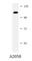
ARG59492 anti-Collagen XVII antibody WB image
Western blot: 20 µg of A2058 cell lysate stained with ARG59492 anti-Collagen XVII antibody at 1:2000 dilution.
-
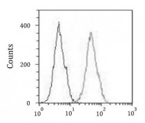
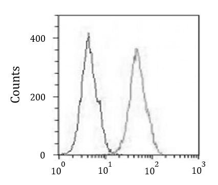
ARG59492 anti-Collagen XVII antibody FACS image
Flow Cytometry: A431 cells were fixed with 2% paraformaldehyde (10 min) and then permeabilized with 90% methanol for 10 min. The cells were then incubated in 2% BSA to block non-specific protein-protein interactions followed by ARG59492 anti-Collagen XVII antibody (right histogram) at 1:25 dilution for 60 min at 37°C, followed by DyLight®488 labelled secondary antibody. Isotype control antibody (left histogram) was rabbit IgG (1 µg/10^6 cells) used under the same conditions. Acquisition of > 10000 events was performed.