ARG57505
anti-Calnexin antibody [18B9]
anti-Calnexin antibody [18B9] for Flow cytometry,ICC/IF,Western blot and Human
概述
| 产品描述 | Mouse Monoclonal antibody [18B9] recognizes Calnexin |
|---|---|
| 反应物种 | Hu |
| 应用 | FACS, ICC/IF, WB |
| 宿主 | Mouse |
| 克隆 | Monoclonal |
| 克隆号 | 18B9 |
| 同位型 | IgG1, kappa |
| 靶点名称 | Calnexin |
| 抗原物种 | Human |
| 抗原 | Recombinant Human Calnexin (aa. 21-481) purified from E. coli. |
| 偶联标记 | Un-conjugated |
| 別名 | P90; CNX; p90; Major histocompatibility complex class I antigen-binding protein p88; Calnexin; IP90 |
应用说明
| 应用建议 |
|
||||||||
|---|---|---|---|---|---|---|---|---|---|
| 应用说明 | * The dilutions indicate recommended starting dilutions and the optimal dilutions or concentrations should be determined by the scientist. |
属性
| 形式 | Liquid |
|---|---|
| 纯化 | Purification with Protein A. |
| 缓冲液 | PBS (pH 7.4), 0.02% Sodium azide and 10% Glycerol. |
| 抗菌剂 | 0.02% Sodium azide |
| 稳定剂 | 10% Glycerol |
| 浓度 | 1 mg/ml |
| 存放说明 | For continuous use, store undiluted antibody at 2-8°C for up to a week. For long-term storage, aliquot and store at -20°C. Storage in frost free freezers is not recommended. Avoid repeated freeze/thaw cycles. Suggest spin the vial prior to opening. The antibody solution should be gently mixed before use. |
| 注意事项 | For laboratory research only, not for drug, diagnostic or other use. |
生物信息
| 数据库连接 | |
|---|---|
| 基因名称 | CANX |
| 全名 | calnexin |
| 背景介绍 | This gene encodes a member of the calnexin family of molecular chaperones. The encoded protein is a calcium-binding, endoplasmic reticulum (ER)-associated protein that interacts transiently with newly synthesized N-linked glycoproteins, facilitating protein folding and assembly. It may also play a central role in the quality control of protein folding by retaining incorrectly folded protein subunits within the ER for degradation. Alternatively spliced transcript variants encoding the same protein have been described. [provided by RefSeq, Jul 2008] |
| 生物功能 | Calcium-binding protein that interacts with newly synthesized glycoproteins in the endoplasmic reticulum. It may act in assisting protein assembly and/or in the retention within the ER of unassembled protein subunits. It seems to play a major role in the quality control apparatus of the ER by the retention of incorrectly folded proteins. Associated with partial T-cell antigen receptor complexes that escape the ER of immature thymocytes, it may function as a signaling complex regulating thymocyte maturation. Additionally it may play a role in receptor-mediated endocytosis at the synapse. [UniProt] |
| 预测分子量 | 68 kDa |
| 翻译后修饰 | Phosphorylated at Ser-564 by MAPK3/ERK1. phosphorylation by MAPK3/ERK1 increases its association with ribosomes (By similarity). Palmitoylation by DHHC6 leads to the preferential localization to the perinuclear rough ER. It mediates the association of calnexin with the ribosome-translocon complex (RTC) which is required for efficient folding of glycosylated proteins. Ubiquitinated, leading to proteasomal degradation. Probably ubiquitinated by ZNRF4. |
检测图片 (4) Click the Picture to Zoom In
-

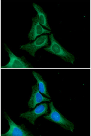
ARG57505 anti-Calnexin antibody [18B9] ICC/IF image
Immunoflorescense: HeLa cells stained with ARG57505 anti-Calnexin antibody [18B9] at 1:100 dilution. The secondary antibody (green) was used Alexa Fluor 488. DAPI was stained the cell nucleus (blue).
-

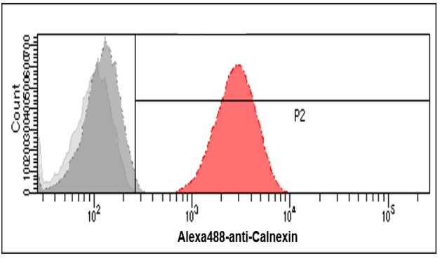
ARG57505 anti-Calnexin antibody [18B9] FACS image
Flow Cytometry: HeLa cells stained with ARG57505 anti-Calnexin antibody [18B9] at 2 - 5 µg/10^6 cells (red). Goat-anti Mouse IgG (Alexa fluor 488) was used as the secondary antibody. Mouse monoclonal IgG was used as the isotype control (dark gray), cells without incubation with primary and secondary antibody was used as the negative control (light gray).
-
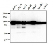
ARG57505 anti-Calnexin antibody [18B9] WB image
Western blot: 40 µg of HeLa, MCF7, A431, A549, 293T, HepG2 and Jurkat cell lysates stained with ARG57505 anti-Calnexin antibody [18B9] at 1:1000 dilution.
-
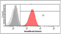
ARG57505 anti-Calnexin antibody [18B9] FACS image
Flow Cytometry: A549 cells stained with ARG57505 anti-Calnexin antibody [18B9] at 2 - 5 µg/10^6 cells (red). Goat-anti Mouse IgG (Alexa fluor 488) was used as the secondary antibody. Mouse monoclonal IgG was used as the isotype control (dark gray), cells without incubation with primary and secondary antibody was used as the negative control (light gray).