ARG10754
anti-CNPase antibody
anti-CNPase antibody for ICC/IF,IHC-Frozen sections,Western blot and Human,Mouse,Rat,Cow
概述
| 产品描述 | Chicken Polyclonal antibody to CNP |
|---|---|
| 反应物种 | Hu, Ms, Rat, Cow |
| 应用 | ICC/IF, IHC-Fr, WB |
| 宿主 | Chicken |
| 克隆 | Polyclonal |
| 同位型 | IgY |
| 靶点名称 | CNPase |
| 抗原物种 | Human |
| 抗原 | Full length Human recombinant protein |
| 偶联标记 | Un-conjugated |
| 別名 | CNPase; 2',3'-cyclic-nucleotide 3'-phosphodiesterase; CNP1; CNP; EC 3.1.4.37 |
应用说明
| 应用建议 |
|
||||||||
|---|---|---|---|---|---|---|---|---|---|
| 应用说明 | * The dilutions indicate recommended starting dilutions and the optimal dilutions or concentrations should be determined by the scientist. |
属性
| 形式 | Liquid |
|---|---|
| 缓冲液 | PBS and 0.02% Sodium azide. |
| 抗菌剂 | 0.02% Sodium azide |
| 存放说明 | For continuous use, store undiluted antibody at 2-8°C for up to a week. For long-term storage, aliquot and store at -20°C or below. Storage in frost free freezers is not recommended. Avoid repeated freeze/thaw cycles. Suggest spin the vial prior to opening. The antibody solution should be gently mixed before use. |
| 注意事项 | For laboratory research only, not for drug, diagnostic or other use. |
生物信息
| 数据库连接 | |
|---|---|
| 基因名称 | CNP |
| 全名 | 2',3'-cyclic nucleotide 3' phosphodiesterase |
| 生物功能 | May participate in RNA metabolism in the myelinating cell, CNP is the third most abundant protein in central nervous system myelin. [UniProt] |
| 预测分子量 | 48 kDa |
检测图片 (4) Click the Picture to Zoom In
-

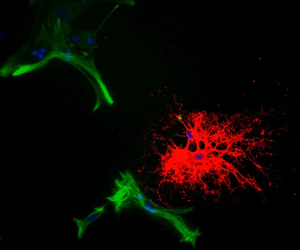
ARG10754 anti-CNPase antibody ICC/IF image
Immunocytochemistry: Rat mixed neuron-glial cell cultures stained with ARG10754 anti-CNPase antibody (red) and co-stained with a Monoclonal 5C10 against GFAP (green). The CNP antibody stains strongly in oligodendrocytes, whereas GFAP labels the intermediate filament in astrocytes. Blue is DNA staining showing the nuclei of these and numerous other cells, most of which are neurons and which do not express either CNP or GFAP.
-

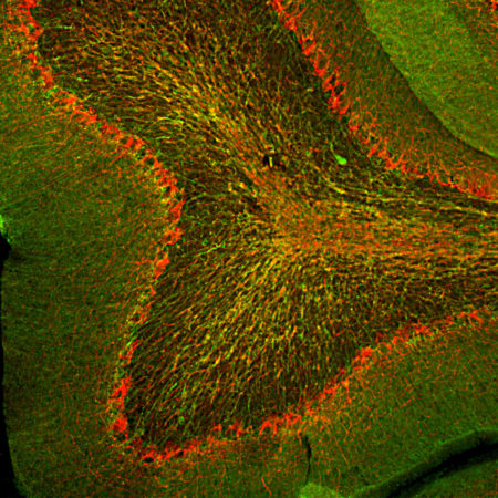
ARG10754 anti-CNPase antibody IHC-Fr image
Immunohistochemistry: Frozen section of Rat cerebellum stained with ARG10754 anti-CNPase antibody (green) at 1:2000 dilution and costained with ARG10761 anti-Neurofilament NF-H antibody (red) at 1:10000 dilution. (Sample preparation: Following transcardial perfusion of Rat with 4% paraformaldehyde, brain was post fixed for 24 hours, cut to 45 µM, and free-floating sections were stained with above antibodies.)
The CNPase antibody stains myelin and oligodendrocytes, cells that create the myelin sheath around axons. The NF-H antibody labels the heavily phosphorylated axonal forms of NF-H which are localized in large projection axons.
-

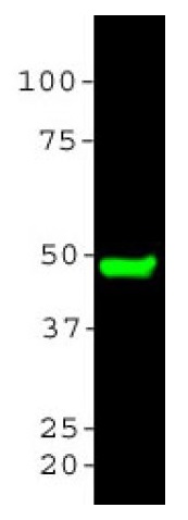
ARG10754 anti-CNPase antibody WB image
Western blot: Rat brain tissue homogenates stained with ARG10754 anti-CNPase antibody at 1:5000.
-

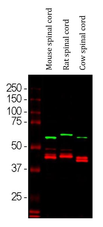
ARG10754 anti-CNPase antibody WB image
Western blot: Mouse spinal cord, Rat spinal cord and Cow spinal cord lysates stained with ARG10754 anti-CNPase antibody (red) at 1:5000 dilution. The blot was simultaneously stained with Mouse mAb to alpha Internexin (green) at 1:2000 dilution.