ARG55775
anti-CLN3 antibody
anti-CLN3 antibody for Flow cytometry,IHC-Formalin-fixed paraffin-embedded sections,Western blot and Human,Mouse
概述
| 产品描述 | Rabbit Polyclonal antibody recognizes CLN3 |
|---|---|
| 反应物种 | Hu, Ms |
| 应用 | FACS, IHC-P, WB |
| 宿主 | Rabbit |
| 克隆 | Polyclonal |
| 同位型 | IgG |
| 靶点名称 | CLN3 |
| 抗原物种 | Human |
| 抗原 | KLH-conjugated synthetic peptide corresponding to aa. 250-284 (Center) of Human CLN3. |
| 偶联标记 | Un-conjugated |
| 別名 | Protein CLN3; JNCL; Batten disease protein; BTS; Battenin |
应用说明
| 应用建议 |
|
||||||||
|---|---|---|---|---|---|---|---|---|---|
| 应用说明 | * The dilutions indicate recommended starting dilutions and the optimal dilutions or concentrations should be determined by the scientist. | ||||||||
| 阳性对照 | SH-SY5Y |
属性
| 形式 | Liquid |
|---|---|
| 纯化 | Purification with Protein A and immunogen peptide. |
| 缓冲液 | PBS and 0.09% (W/V) sodium azide. |
| 抗菌剂 | 0.09% (W/V) sodium azide. |
| 存放说明 | For continuous use, store undiluted antibody at 2-8°C for up to a week. For long-term storage, aliquot and store at -20°C or below. Storage in frost free freezers is not recommended. Avoid repeated freeze/thaw cycles. Suggest spin the vial prior to opening. The antibody solution should be gently mixed before use. |
| 注意事项 | For laboratory research only, not for drug, diagnostic or other use. |
生物信息
| 数据库连接 | |
|---|---|
| 基因名称 | CLN3 |
| 全名 | ceroid-lipofuscinosis, neuronal 3 |
| 背景介绍 | This gene encodes a protein that is involved in lysosomal function. Mutations in this, as well as other neuronal ceroid-lipofuscinosis (CLN) genes, cause neurodegenerative diseases commonly known as Batten disease or collectively known as neuronal ceroid lipofuscinoses (NCLs). Many alternatively spliced transcript variants have been found for this gene. [provided by RefSeq, Jul 2008] |
| 生物功能 | Involved in microtubule-dependent, anterograde transport of late endosomes and lysosomes. [UniProt] |
| 细胞定位 | Lysosome membrane; Multi- pass membrane protein. Late endosome |
| 预测分子量 | 48 kDa |
| 翻译后修饰 | Highly glycosylated. Farnesylation is important for trafficking to lysosomes. |
检测图片 (3) Click the Picture to Zoom In
-
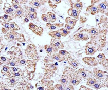
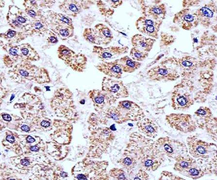
ARG55775 anti-CLN3 antibody IHC-P image
Immunohistochemistry: Paraffin-embedded Human liver tissue stained with ARG55775 anti-CLN3 antibody at 1:25 dilution.
-
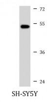
ARG55775 anti-CLN3 antibody WB image
Western blot: 20 µg of SH-SY5Y cell lysate stained with ARG55775 anti-CLN3 antibody at 1:1000 dilution.
-
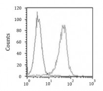
ARG55775 anti-CLN3 antibody FACS image
Flow Cytometry: HeLa cells stained with ARG55775 anti-CLN3 antibody (right histogram) at 1:25 dilution or isotype control antibody (left histogram), followed by incubation with Alexa Fluor® 488 labelled secondary antibody.