ARG66999
anti-CD279 / PD-1 antibody
anti-CD279 / PD-1 antibody for Flow cytometry,IHC-Formalin-fixed paraffin-embedded sections,Western blot and Human
概述
| 产品描述 | Mouse Monoclonal antibody recognizes CD279 / PD-1 |
|---|---|
| 反应物种 | Hu |
| 预测物种 | Ms, Rat |
| 应用 | FACS, IHC-P, WB |
| 宿主 | Mouse |
| 克隆 | Monoclonal |
| 同位型 | IgG2a |
| 靶点名称 | CD279 / PD-1 |
| 抗原物种 | Human |
| 抗原 | Recombinant protein corresponding to Human PD-1. |
| 偶联标记 | Un-conjugated |
| 別名 | hPD-l; CD279; PD-1; Protein PD-1; CD antigen CD279; PD1; hSLE1; SLEB2; Programmed cell death protein 1; hPD-1 |
应用说明
| 应用建议 |
|
||||||||
|---|---|---|---|---|---|---|---|---|---|
| 应用说明 | * The dilutions indicate recommended starting dilutions and the optimal dilutions or concentrations should be determined by the scientist. | ||||||||
| 阳性对照 | Smooth muscle tissue; anti-CD3, anti-CD28 actived PBMC | ||||||||
| 实际分子量 | 55 kDa |
属性
| 形式 | Liquid |
|---|---|
| 纯化 | Affinity purified. |
| 缓冲液 | PBS (pH 7.2) and 0.09% Sodium azide. |
| 抗菌剂 | 0.09% Sodium azide |
| 存放说明 | For continuous use, store undiluted antibody at 2-8°C for up to a week. For long-term storage, aliquot and store at -20°C or below. Storage in frost free freezers is not recommended. Avoid repeated freeze/thaw cycles. Suggest spin the vial prior to opening. The antibody solution should be gently mixed before use. |
| 注意事项 | For laboratory research only, not for drug, diagnostic or other use. |
生物信息
| 数据库连接 | |
|---|---|
| 基因名称 | PDCD1 |
| 全名 | programmed cell death 1 |
| 背景介绍 | CD279 / PD-1 is a cell surface membrane protein of the immunoglobulin superfamily. This protein is expressed in pro-B-cells and is thought to play a role in their differentiation. In mice, expression of this gene is induced in the thymus when anti-CD3 antibodies are injected and large numbers of thymocytes undergo apoptosis. Mice deficient for this gene bred on a BALB/c background developed dilated cardiomyopathy and died from congestive heart failure. These studies suggest that this gene product may also be important in T cell function and contribute to the prevention of autoimmune diseases. [provided by RefSeq, Jul 2008] |
| 生物功能 | CD279 / PD-1 is an inhibitory receptor on antigen activated T-cells. It plays a critical role in induction and maintenance of immune tolerance to self (PubMed:21276005). Delivers inhibitory signals upon binding to ligands CD274/PDCD1L1 and CD273/PDCD1LG2 (PubMed:21276005). Following T-cell receptor (TCR) engagement, PDCD1 associates with CD3-TCR in the immunological synapse and directly inhibits T-cell activation. Suppresses T-cell activation through the recruitment of PTPN11/SHP-2: following ligand-binding, PDCD1 is phosphorylated within the ITSM motif, leading to the recruitment of the protein tyrosine phosphatase PTPN11/SHP-2 that mediates dephosphorylation of key TCR proximal signaling molecules, such as ZAP70, PRKCQ/PKCtheta and CD247/CD3zeta. The PDCD1-mediated inhibitory pathway is exploited by tumors to attenuate anti-tumor immunity and escape destruction by the immune system, thereby facilitating tumor survival (PubMed:28951311). The interaction with CD274/PDCD1L1 inhibits cytotoxic T lymphocytes (CTLs) effector function (PubMed:28951311). The blockage of the PDCD1-mediated pathway results in the reversal of the exhausted T-cell phenotype and the normalization of the anti-tumor response, providing a rationale for cancer immunotherapy (PubMed:22658127, PubMed:25034862, PubMed:25399552). [UniProt] |
| 细胞定位 | Membrane |
| 产品亮点 | Related products: PD-1 antibodies; PD-1 ELISA Kits; PD-1 Duos / Panels; Anti-Mouse IgG secondary antibodies; Related news: New PD-1 ELISA Kit, excellent for preclinical studies or pharmatheutical development Why anti-PD-1/PD-L1 therapy doesn’t work? The best solution for PD-1/PD-L1 research Examining CTL/NK-mediated cytotoxicity by ELISA |
| 预测分子量 | 32 kDa |
检测图片 (2) Click the Picture to Zoom In
-

ARG66999 anti-CD279 / PD-1 antibody IHC-P image
Immunohistochemistry: Paraffin-embedded Human smooth muscle tissue stained with ARG66999 anti-CD279 / PD-1 antibody. Antigen Retrieval: Boil tissue section in Citrate buffer (pH 6.0).
-
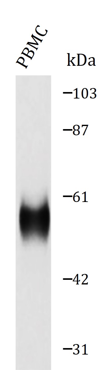
ARG66999 anti-CD279 / PD-1 antibody WB image
Western blot: Lysate from anti-CD3 and antiCD28 actived PBMC cell stained with ARG66999 anti-CD279 / PD-1 antibody, overnight at 4°C.