ARG66321
anti-CD19 antibody [SQab1869]
anti-CD19 antibody [SQab1869] for Flow cytometry,ICC/IF,IHC-Formalin-fixed paraffin-embedded sections,Immunoprecipitation,Western blot and Human
Developmental Biology antibody; Immune System antibody; Lymphocyte Marker antibody; B cell Marker antibody; Pro-B Cell Marker antibody; Pre-B Cell Marker antibody; Immature B Cell Marker antibody; Follicular dendritic cells antibody
概述
| 产品描述 | Recombinant Rabbit Monoclonal antibody [SQab1869] recognizes CD19 |
|---|---|
| 反应物种 | Hu |
| 应用 | FACS, ICC/IF, IHC-P, IP, WB |
| 宿主 | Rabbit |
| 克隆 | Monoclonal |
| 克隆号 | SQab1869 |
| 同位型 | IgG |
| 靶点名称 | CD19 |
| 抗原物种 | Human |
| 抗原 | Synthetic peptide around the C-terminus of CD19. |
| 偶联标记 | Un-conjugated |
| 別名 | Differentiation antigen CD19; T-cell surface antigen Leu-12; B-lymphocyte antigen CD19; B-lymphocyte surface antigen B4; B4; CD antigen CD19; CVID3 |
应用说明
| 应用建议 |
|
||||||||||||
|---|---|---|---|---|---|---|---|---|---|---|---|---|---|
| 应用说明 | IHC-P: Antigen Retrieval: Heat mediated was performed using Tris/EDTA buffer pH 9.0. * The dilutions indicate recommended starting dilutions and the optimal dilutions or concentrations should be determined by the scientist. |
属性
| 形式 | Liquid |
|---|---|
| 纯化 | Purification with Protein A. |
| 缓冲液 | PBS, 0.01% Sodium azide, 40% Glycerol and 0.05% BSA. |
| 抗菌剂 | 0.01% Sodium azide |
| 稳定剂 | 40% Glycerol and 0.05% BSA |
| 存放说明 | For continuous use, store undiluted antibody at 2-8°C for up to a week. For long-term storage, aliquot and store at -20°C. Storage in frost free freezers is not recommended. Avoid repeated freeze/thaw cycles. Suggest spin the vial prior to opening. The antibody solution should be gently mixed before use. |
| 注意事项 | For laboratory research only, not for drug, diagnostic or other use. |
生物信息
| 数据库连接 | |
|---|---|
| 基因名称 | CD19 |
| 全名 | CD19 molecule |
| 背景介绍 | CD19: Lymphocytes proliferate and differentiate in response to various concentrations of different antigens. The ability of the B cell to respond in a specific, yet sensitive manner to the various antigens is achieved with the use of low-affinity antigen receptors. This gene encodes a cell surface molecule which assembles with the antigen receptor of B lymphocytes in order to decrease the threshold for antigen receptor-dependent stimulation. [provided by RefSeq, Jul 2008] |
| 生物功能 | CD19 functions as coreceptor for the B-cell antigen receptor complex (BCR) on B-lymphocytes. Decreases the threshold for activation of downstream signaling pathways and for triggering B-cell responses to antigens (PubMed:2463100, PubMed:1373518, PubMed:16672701). Activates signaling pathways that lead to the activation of phosphatidylinositol 3-kinase and the mobilization of intracellular Ca(2+) stores (PubMed:9382888, PubMed:9317126, PubMed:12387743, PubMed:16672701). Is not required for early steps during B cell differentiation in the blood marrow (PubMed:9317126). Required for normal differentiation of B-1 cells. Required for normal B cell differentiation and proliferation in response to antigen challenges (PubMed:2463100, PubMed:1373518). Required for normal levels of serum immunoglobulins, and for production of high-affinity antibodies in response to antigen challenge (PubMed:9317126, PubMed:12387743, PubMed:16672701). [UniProt] |
| 产品亮点 | Related news: Cancer Pathology Markers (SQ clones); Tumor-Infiltrating Lymphocytes (TILs); MyD88 L265P antibody for lymphoma research; Related products: CD19 antibodies; CD19 ELISA Kits; CD19 Duos / Panels; Anti-Rabbit IgG secondary antibodies; |
| 研究领域 | Developmental Biology antibody; Immune System antibody; Lymphocyte Marker antibody; B cell Marker antibody; Pro-B Cell Marker antibody; Pre-B Cell Marker antibody; Immature B Cell Marker antibody; Follicular dendritic cells antibody |
| 预测分子量 | 61 kDa |
| 翻译后修饰 | Phosphorylated on serine and threonine upon DNA damage, probably by ATM or ATR. Phosphorylated on tyrosine following B-cell activation. Phosphorylated on tyrosine residues by LYN. [UniProt] |
检测图片 (5) Click the Picture to Zoom In
-
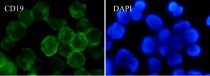
ARG66321 anti-CD19 antibody [SQab1869] ICC/IF image
Immunofluorescence: Raji cells were fixed with 4% paraformaldehyde for 30 min at RT, permeabilized with 0.1% Triton X-100 for 10 min at RT then blocked with 10% goat serum for 30 min at RT. Cells were stained with ARG66321 anti-CD19 antibody [SQab1869] (green) at 1:50 and 4°C. DAPI (blue) was used as the nuclear counter stain.
-
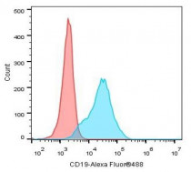
ARG66321 anti-CD19 antibody [SQab1869] FACS image
Flow Cytometry: Ramos cells were fixed with 4% paraformaldehyde for 10 mins. The cells were stained with ARG66321 anti-CD19 antibody [SQab1869] (blue) at 1:10 dilution in 1x PBS/1% BSA for 30 min at RT, followed by Alexa Fluor® 488 labelled secondary antibody. Unlabelled sample (red) was used as a control.
-
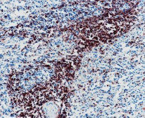
ARG66321 anti-CD19 antibody [SQab1869] IHC-P image
Immunohistochemistry: Formalin-fixed and paraffin-embedded spleen tissue stained with ARG66321 anti-CD19 antibody [SQab1869] at 1:400 dilution. Antigen Retrieval: Heat mediated was performed using Tris/EDTA buffer pH 9.0.
-

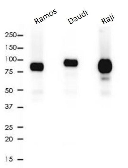
ARG66321 anti-CD19 antibody [SQab1869] WB image
Western blot: 10 µg of Ramos, Daudi and Raji cell lysates stained with ARG66321 anti-CD19 antibody [SQab1869] at 1:2000 dilution.
-
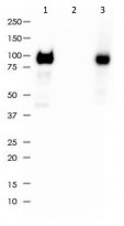
ARG66321 anti-CD19 antibody [SQab1869] IP image
Immunoprecipitation: 0.4 mg of Ramos lysate immunoprecipitated (1:50) and stained with ARG66321 anti-CD19 antibody [SQab1869]. 1) ARG66321 IP in Ramos whole cell lysate, 2) Rabbit IgG instead of ARG66321 in Ramos whole cell lysate, and 3) Ramos whole cell lysate, 10 µg (input).