ARG58313
anti-Bik antibody
anti-Bik antibody for Flow cytometry,IHC-Formalin-fixed paraffin-embedded sections,Western blot and Human,Mouse,Rat
概述
| 产品描述 | Rabbit Polyclonal antibody recognizes Bik |
|---|---|
| 反应物种 | Hu, Ms, Rat |
| 应用 | FACS, IHC-P, WB |
| 宿主 | Rabbit |
| 克隆 | Polyclonal |
| 同位型 | IgG |
| 靶点名称 | Bik |
| 抗原物种 | Human |
| 抗原 | Human Bik recombinant protein (Position: M1-R123). Human Bik shares 42.3% amino acid (aa) sequence identity with Mouse Bik. |
| 偶联标记 | Un-conjugated |
| 別名 | NBK; Bcl-2-interacting killer; BIP1; Apoptosis inducer NBK; BP4 |
应用说明
| 应用建议 |
|
||||||||
|---|---|---|---|---|---|---|---|---|---|
| 应用说明 | IHC-P: Antigen Retrieval: Heat mediated was performed in Citrate buffer (pH 6.0) for 20 min. * The dilutions indicate recommended starting dilutions and the optimal dilutions or concentrations should be determined by the scientist. |
属性
| 形式 | Liquid |
|---|---|
| 纯化 | Affinity purification with immunogen. |
| 缓冲液 | 0.9% NaCl, 0.2% Na2HPO4, 0.05% Sodium azide and 5% BSA. |
| 抗菌剂 | 0.05% Sodium azide |
| 稳定剂 | 5% BSA |
| 浓度 | 0.5 mg/ml |
| 存放说明 | For continuous use, store undiluted antibody at 2-8°C for up to a week. For long-term storage, aliquot and store at -20°C or below. Storage in frost free freezers is not recommended. Avoid repeated freeze/thaw cycles. Suggest spin the vial prior to opening. The antibody solution should be gently mixed before use. |
| 注意事项 | For laboratory research only, not for drug, diagnostic or other use. |
生物信息
| 数据库连接 | |
|---|---|
| 基因名称 | BIK |
| 全名 | BCL2-interacting killer (apoptosis-inducing) |
| 背景介绍 | The protein encoded by this gene shares a critical BH3 domain with other death-promoting proteins, such as BID, BAK, BAD and BAX, that is required for its pro-apoptotic activity, and for interaction with anti-apoptotic members of the BCL2 family, and viral survival-promoting proteins. Since the activity of this protein is suppressed in the presence of survival-promoting proteins, it is suggested as a likely target for anti-apoptotic proteins. [provided by RefSeq, Sep 2011] |
| 生物功能 | Accelerates programmed cell death. Association to the apoptosis repressors Bcl-X(L), BHRF1, Bcl-2 or its adenovirus homolog E1B 19k protein suppresses this death-promoting activity. Does not interact with BAX. [UniProt] |
| 细胞定位 | Endomembrane system; Single-pass membrane protein. Mitochondrion membrane; Single-pass membrane protein. Around the nuclear envelope, and in cytoplasmic membranes. [UniProt] |
| 预测分子量 | 18 kDa |
| 翻译后修饰 | Proteolytically cleaved by RHBDL4/RHBDD1. RHBDL4/RHBDD1-induced cleavage is a necessary step prior its degradation by the proteosome-dependent mechanism. [UniProt] |
检测图片 (7) Click the Picture to Zoom In
-
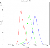
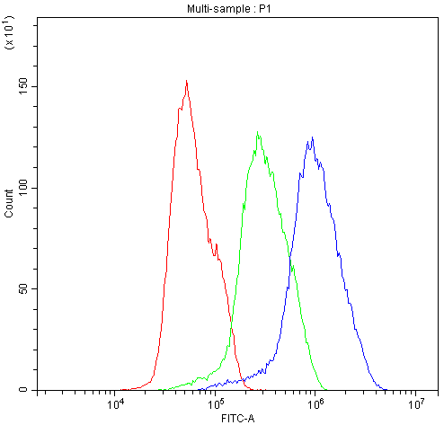
ARG58313 anti-Bik antibody FACS image
Flow Cytometry: MCF-7 cells were blocked with 10% normal goat serum, and then stained with ARG58313 anti-Bik antibody (blue) at 1 µg/10^6 cells for 30 min at 20°C, followed by DyLight®488 labelled secondary antibody. Isotype control antibody (green) was rabbit IgG (1 µg/10^6 cells) used under the same conditions. Unlabelled sample (red) was also used as a control.
-

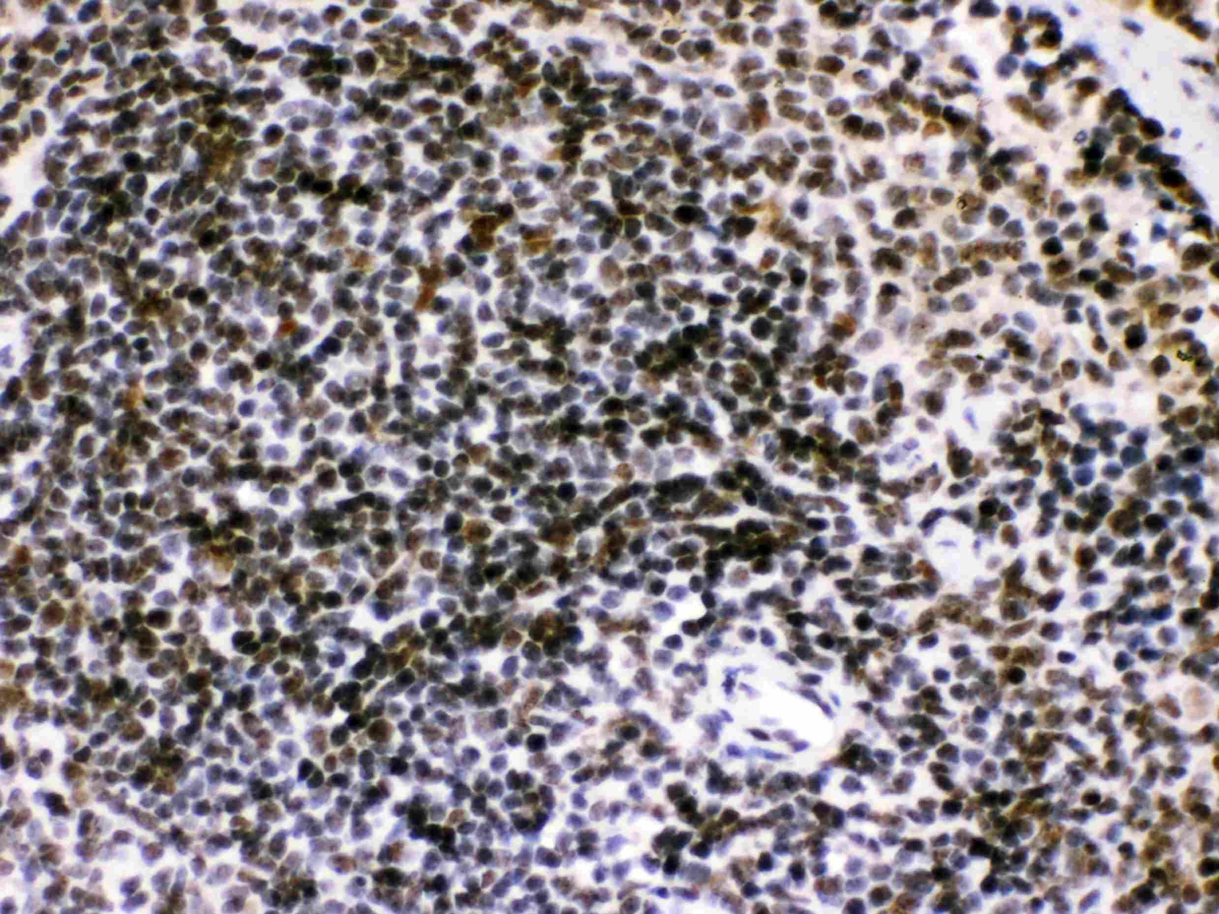
ARG58313 anti-Bik antibody IHC-P image
Immunohistochemistry: Paraffin-embedded Mouse spleen tissue. Antigen Retrieval: Heat mediated was performed in Citrate buffer (pH 6.0, epitope retrieval solution) for 20 min. The tissue section was blocked with 10% goat serum. The tissue section was then stained with ARG58313 anti-Bik antibody at 1 µg/ml dilution, overnight at 4°C.
-
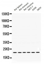
ARG58313 anti-Bik antibody WB image
Western blot: 50 µg of Rat spleen, Rat stomach, Rat intestine, MCF-7 whole cell lysate, A549 whole cell lysate and SKOV whole cell lysate stained with ARG58313 anti-Bik antibody at 0.5 µg/ml, overnight at 4°C, under reducing conditions.
-
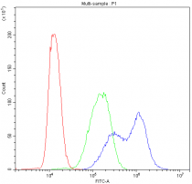
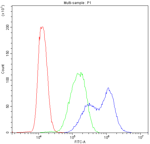
ARG58313 anti-Bik antibody FACS image
Flow Cytometry: THP-1 cells were blocked with 10% normal goat serum, and then stained with ARG58313 anti-Bik antibody (blue) at 1 µg/10^6 cells for 30 min at 20°C, followed by DyLight®488 labelled secondary antibody. Isotype control antibody (green) was rabbit IgG (1 µg/10^6 cells) used under the same conditions. Unlabelled sample (red) was also used as a control.
-
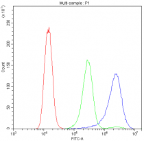
ARG58313 anti-Bik antibody FACS image
Flow Cytometry: A431 cells were blocked with 10% normal goat serum, and then stained with ARG58313 anti-Bik antibody (blue) at 1 µg/10^6 cells for 30 min at 20°C, followed by DyLight®488 labelled secondary antibody. Isotype control antibody (green) was rabbit IgG (1 µg/10^6 cells) used under the same conditions. Unlabelled sample (red) was also used as a control.
-
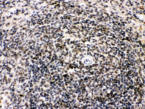
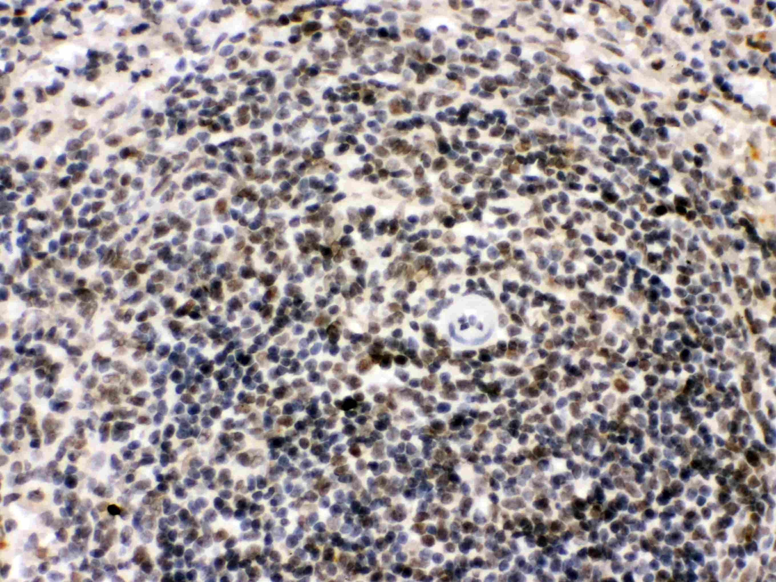
ARG58313 anti-Bik antibody IHC-P image
Immunohistochemistry: Paraffin-embedded Rat spleen tissue. Antigen Retrieval: Heat mediated was performed in Citrate buffer (pH 6.0, epitope retrieval solution) for 20 min. The tissue section was blocked with 10% goat serum. The tissue section was then stained with ARG58313 anti-Bik antibody at 1 µg/ml dilution, overnight at 4°C.
-
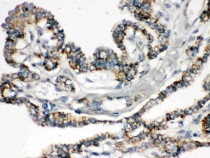
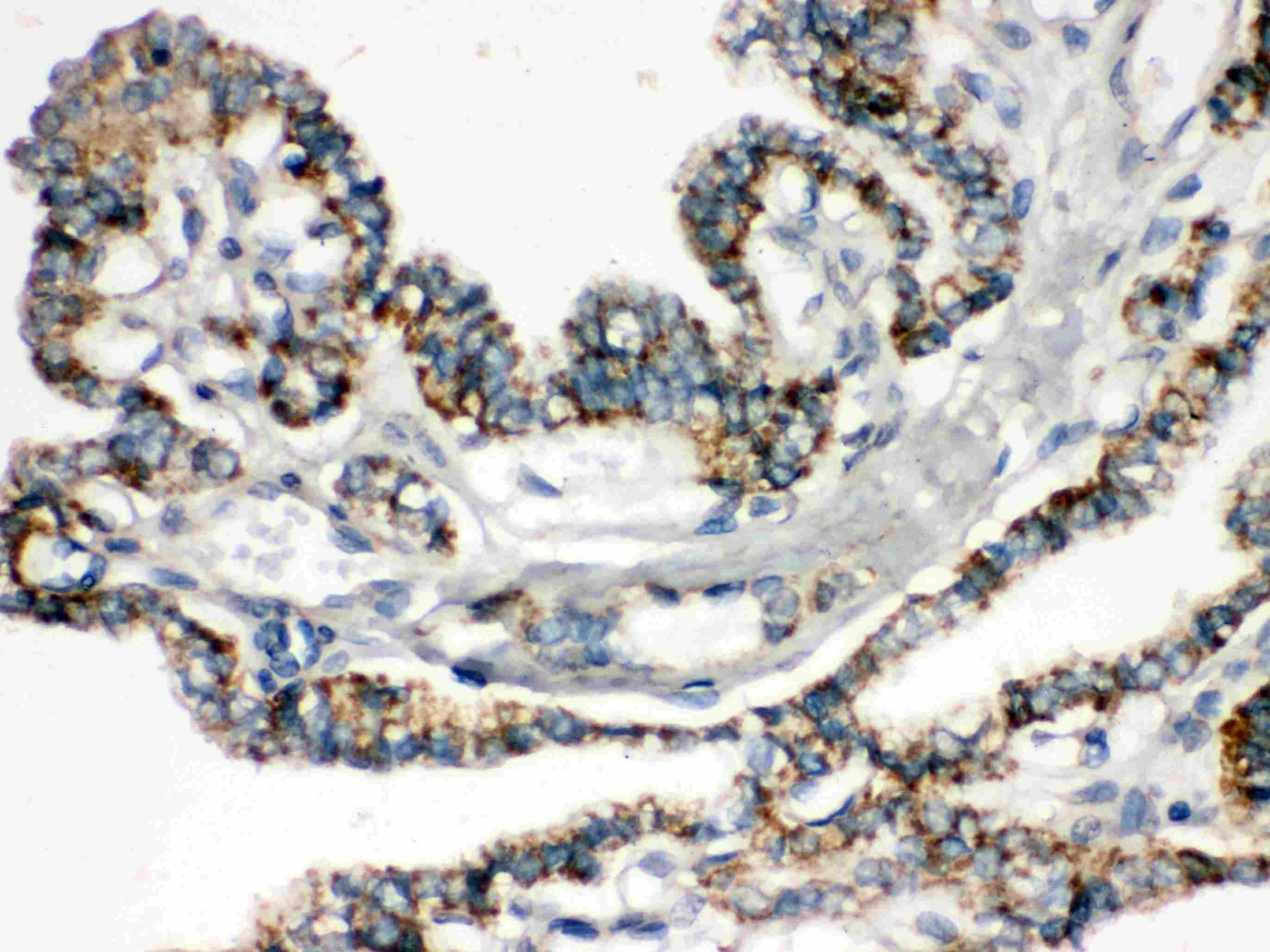
ARG58313 anti-Bik antibody IHC-P image
Immunohistochemistry: Paraffin-embedded Human thyroid cancer tissue. Antigen Retrieval: Heat mediated was performed in Citrate buffer (pH 6.0, epitope retrieval solution) for 20 min. The tissue section was blocked with 10% goat serum. The tissue section was then stained with ARG58313 anti-Bik antibody at 1 µg/ml dilution, overnight at 4°C.